Disclaimer: This article is for informational purposes only and is not intended for diagnostic use. LifeDNA does not provide diagnostic reports on any traits discussed. Genetics is just one piece of the puzzle; please consult a healthcare professional for comprehensive guidance on any health condition.
What Is Mitral Valve Prolapse?
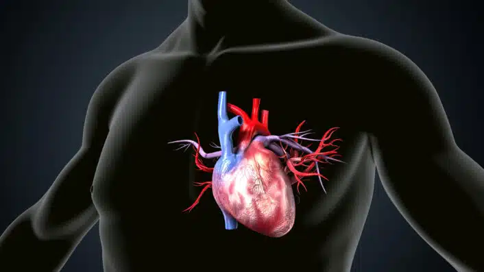
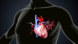
Mitral Valve Prolapse or MVP is a condition where one of the heart’s valves doesn’t close properly. The mitral valve is located between the left atrium and the left ventricle. In MVP, the flaps, also called leaflets, of this valve bulge or prolapse slightly backward into the left atrium when the heart contracts.
For most people, this doesn’t cause any serious health issues. In fact, many people don’t even know they have it until it shows up during a routine check-up. But in some cases, it may lead to complications like mitral regurgitation, when blood leaks backward into the atrium, irregular heartbeats, or rarely, more serious cardiovascular problems.
You may want to read: Unraveling The Genetics of Resting Heart Rate
What Causes Mitral Valve Prolapse?
Mitral valve prolapse or MVP happens when the valve between the heart’s left upper chamber (left atrium) and lower chamber (left ventricle) doesn’t close properly. Instead of closing tightly, the valve’s flaps—called leaflets—become floppy and bulge backward into the upper chamber during a heartbeat. But what causes this to happen? The answer often lies in the structure and strength of connective tissue, which forms the framework of the valve.
The mitral valve is made up of connective tissue that gives it flexibility and strength. Over time, this tissue can become stretched or weakened, which causes the valve leaflets to lose their shape and function. As people age, connective tissues throughout the body—including those in the heart—can gradually break down. This age-related wear and tear can weaken the valve and its supporting structures (like the chordae tendineae), increasing the chances of prolapse.This is especially true in people whose connective tissue is naturally more elastic or prone to damage.
Here are some specific the causes and how they affect the heart:
- Associated medical conditions that affect body structure: Some health conditions, such as scoliosis or curvature of the spine or muscular dystrophy known as a group of genetic disorders that weaken muscles, may influence the way connective tissues develop or function. These changes in the connective tissue can also impact the heart valves, making a prolapse more likely.
- Infections like rheumatic fever: In some people, a past infection like rheumatic fever, which can follow untreated strep throat may cause inflammation and scarring of the heart valves. This damage can permanently alter the valve’s shape and movement, leading to MVP.
- Congenital (present at birth) MVP: Some people are born with slightly abnormal mitral valves due to genetic or developmental factors. In these cases, MVP may not show symptoms early in life but becomes noticeable later on, especially if the valve’s shape changes further with age.
- Other connective tissue disorders: Certain inherited conditions such as Marfan syndrome or Ehlers-Danlos syndrome directly affect the strength and elasticity of connective tissue. These disorders can lead to floppy mitral valve leaflets and stretchy chordae tendineae, increasing the risk of MVP as part of a broader syndrome that may affect multiple organs and systems.
- Lifestyle and environmental factors: Although less common as direct causes, factors like chronic high blood pressure, poor diet, lack of exercise, or long-term stress may indirectly contribute to valve weakening. These factors place extra strain on the heart over time, which may lead to structural changes, especially in people who are already genetically predisposed to MVP.
MVP can be a a result from a mix of genetic factors (see below), physical stress on the heart, and diseases that affect the body’s connective tissues. In many cases, the cause is a combination of inherited tissue characteristics and age-related changes.
Common Symptoms of Mitral Valve Prolapse
Many people with mitral valve prolapse (MVP) don’t notice any symptoms at all. But for those who do, the signs can vary from mild to more bothersome. They may also come and go over time. Here’s a closer look at what some of these symptoms might feel like:
- Heart palpitations: You might feel like your heart is fluttering, pounding, or skipping beats. These unusual rhythms can be brief or last for several minutes, and they often happen even when you’re resting.
- Chest discomfort: This isn’t usually a sharp pain. Instead, it may feel like a dull pressure, tightness, or an aching sensation in your chest. It can be unsettling, but it doesn’t always mean there’s a serious problem.
- Fatigue: Feeling unusually tired or drained, even after a full night’s sleep or without doing much physical activity, is a common symptom. This kind of fatigue may feel different from typical tiredness.
- Dizziness or lightheadedness: Some people feel unsteady, faint, or like the room is spinning. This can happen when standing up quickly or during periods of stress or exertion.
- Shortness of breath: You may notice it’s harder to catch your breath, especially when you’re exercising, walking up stairs, or even lying flat. This can happen because the heart isn’t pumping blood as efficiently as it should.
These symptoms can vary from person to person and may not always be linked directly to MVP. However, if you experience them frequently or if they get worse over time, it’s important to talk to your healthcare provider. Keeping track of your symptoms can help guide proper diagnosis and treatment.
Is Mitral Valve Prolapse Genetic?
Studies suggest that mitral valve prolapse has a genetic component. While MVP can happen on its own, researchers have found that it can run in families, showing that genes play an important role.
One of the earliest signs of a genetic link was seen in families where several members had MVP. Some early cases suggested the condition might be passed down through the X chromosome, but later studies showed that autosomal dominant inheritance is more common. This means that if one parent has the genetic factor linked to MVP, their children have about a 50% chance of inheriting it.
There are different types of MVP based on how the valve tissue is affected. One type called myxomatous MVP, also known as Barlow’s disease, tends to happen in younger people and is strongly linked to inherited gene variants. Another type, called fibroelastic deficiency (FED), usually appears later in life and is more related to aging than genetics.
Several genes have been identified in relation to MVP, some playing a role especially in families with inherited forms. These include:
- FLNA : This gene is linked to a rare X-linked form of MVP and can cause serious valve problems.
- DCHS1: This gene helps with the structure and organization of heart cells, playing a role in how the valve develops.
- DZIP1: This gene supports the function of cellular structures important for heart valve formation.
However, these rare genetic mutations only explain a small portion of MVP cases. Most people with MVP likely have common genetic variants, SNPs, which are tiny changes in their DNA that each slightly raise their risk of developing the condition. These were discovered through genome-wide association studies (GWAS), a type of research that looks at the entire genome to find patterns linked to disease.
What is interesting is that some family members may not show full-blown MVP, but instead may have early or subtle signs, like slight changes in valve structure. These early signs may still be genetically influenced and may develop into full MVP over time. In fact, studies have shown that children are more likely to develop MVP if their parents have even mild valve changes.
Researchers also think there may be a genetic link between MVP and other heart problems, such as ventricular arrhythmia or certain forms of cardiomyopathy. This suggests that MVP may be a part of a bigger genetic pattern affecting the heart’s structure and rhythm.
To understand these connections better, scientists use animal models. These models are bred to carry the same genetic changes found in humans with MVP. Studying how these changes affect heart structure and function helps researchers uncover the underlying causes and possible ways to treat or prevent MVP. Genetics plays a major role in MVP, especially in cases that run in families or appear at a young age. Both rare mutations and more common genetic traits contribute to how and when MVP develops. Recognizing this genetic connection is key to improving diagnosis, guiding family screening, and eventually finding better treatments.
How Is Mitral Valve Prolapse Diagnosed?
Mitral Valve Prolapse is often discovered during a routine physical exam. Your doctor might hear a distinctive clicking sound or murmur with a stethoscope, which can lead to further tests. If mitral valve prolapse or MVP is suspected, your doctor may order several tests to confirm the diagnosis and understand how well your heart is functioning. These tests can provide a clearer picture of your heart’s structure and rhythm. Common diagnostic tools include:
- Chest X-ray or Cardiac MRI: These imaging tests are used in certain cases to view the size, shape, and overall structure of your heart. A chest X-ray can show if the heart is enlarged, while a cardiac MRI provides highly detailed images of the heart’s anatomy and function.
- Echocardiogram: This is the most accurate and commonly used test to confirm MVP. It uses sound waves or ultrasound to create detailed images of your heart. Doctors can see the mitral valve’s leaflets in motion and check if they are bulging backward or leaking. With advanced 3D echocardiography, the images are even more precise, helping doctors assess the severity of the condition in real-time.
- Electrocardiogram: This test records your heart’s electrical activity. It helps detect abnormal rhythms or arrhythmias that sometimes occur with MVP, especially if you’re experiencing palpitations or dizziness.
Is Mitral Valve Prolapse Dangerous?
Mitral valve prolapse (MVP) is usually not dangerous. Most people with this condition live normal, healthy lives without any serious problems. However, in some cases, MVP can lead to complications—especially if the valve’s flaps are more severely affected. The most common complication is mitral regurgitation, which happens when the valve doesn’t close properly and allows blood to leak backward into the upper chamber of the heart. This leakage can put extra strain on the heart and may cause symptoms like tiredness, shortness of breath, or reduced ability to exercise.
Another possible complication is arrhythmia, which means the heart beats irregularly. Some people may feel heart palpitations, fluttering, or even dizziness. While most arrhythmias are harmless, some may require treatment. A rare but serious risk is endocarditis, an infection of the heart’s inner lining or valves. People with damaged valves are more likely to develop this infection, which can be life-threatening and usually requires strong antibiotics or even surgery. In very rare cases, severe mitral valve damage can lead to heart failure. This happens when the heart can no longer pump blood effectively. Regular checkups and early treatment can help prevent these complications and keep the heart working well.
For many, it’s a manageable condition that doesn’t interfere with daily life. However, if you experience symptoms or are at higher risk of complications, there are several ways to take care of your heart and improve your quality of life.
- Regular heart monitoring: Periodic checkups with tests like echocardiograms (ultrasound images of the heart) or electrocardiograms (ECGs) help track how well your mitral valve is working. These tests can catch early signs of valve changes or irregular heart rhythms, even before symptoms appear.
- Medications if needed: Some people may be prescribed beta-blockers or other medications to help manage symptoms such as heart palpitations or to treat high blood pressure. These drugs work by slowing the heart rate and reducing the heart’s workload, helping you feel more comfortable and reducing your risk of complications.
- Staying active with moderation: Physical activity is generally safe and encouraged for people with MVP, as long as it’s not too intense. Moderate exercise, like walking, swimming, or cycling, helps strengthen the heart and improve overall well-being. However, overexertion or intense endurance activities might not be suitable for everyone—your doctor can help you find the right balance.
- Avoiding stimulants: Substances like caffeine, nicotine, or certain cold medications can sometimes make heart palpitations worse. Limiting or avoiding these stimulants may reduce uncomfortable symptoms and help keep your heartbeat more steady.
- Managing stress: Emotional stress and anxiety can increase symptoms like chest discomfort or palpitations. Stress-reducing practices such as mindfulness, deep breathing, yoga, or cognitive behavioral therapy (CBT) can be especially helpful in improving both mental and heart health.
References