Are Varicose Veins Genetic?

What Are Varicose Veins?

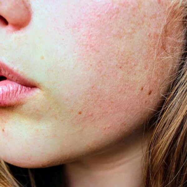
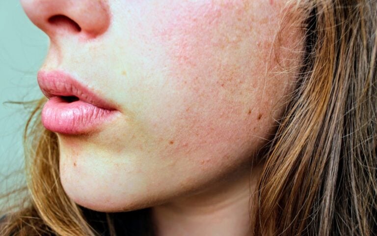
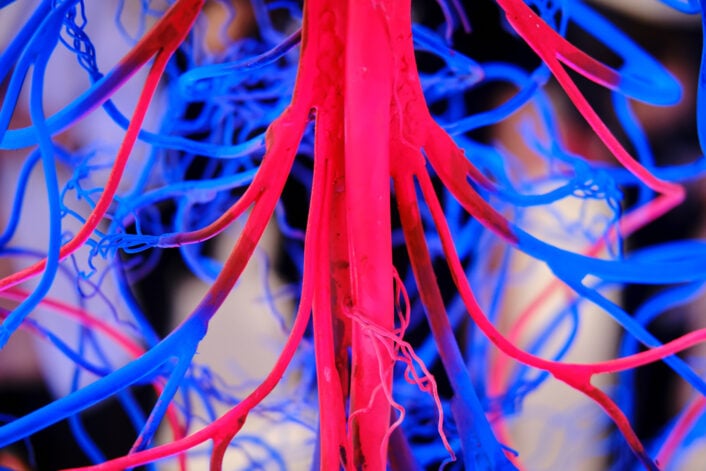
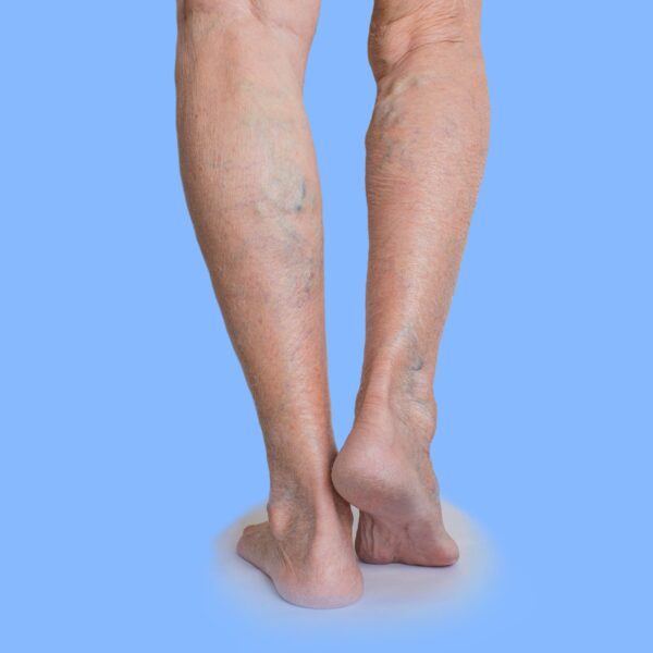
Varicose veins are a common condition of the blood vessels characterized by swollen, enlarged, and twisted veins visible beneath the skin’s surface. Most frequently occurring in the legs and feet, these veins can range from dark purple to blue. Though varicose veins can be unsightly, they often pose more than just a cosmetic problem; they can lead to discomfort, pain, and sometimes severe medical issues related to the circulatory system.

A healthy vein allows blood to flow smoothly back to the heart through a series of one-way valves. In a varicose vein, these valves malfunction, allowing blood to accumulate, leading to enlargement and a twisted, rope-like appearance. While smaller veins are often red or purple, more prominent varicose veins tend to be bluish.

Are Varicose Veins Genetic?

While varicose veins can be influenced by factors like age, weight, and lifestyle, there could also be a genetic component.

Research on Genetic Markers

Studies on the genetic aspects of varicose veins have mainly focused on symptoms of specific disorders of the blood vessels (like Klippel-Trenaunay Syndrome). Previous research indicates that changes in genes like FOXC2, thrombomodulin (THBD), and desmuslin (SYNM) could contribute to varicose veins by affecting how veins function. However, these investigations have generally involved small sample sizes, ranging from 18 to 700 participants, and there needs to be more follow-up studies to confirm these findings.

A 2019 Genome-Wide Association Study (GWAS), including nearly 10,000 cases and 300,000 controls, identified 30 genetic locations strongly linked with varicose veins. The most notable associations were found in the intron region of CASZ1 (rs1112165), previously implicated in blood pressure, and in the 16q24 region, where the PIEZO1 gene is located.

CASZ1 Gene

In the past few years, multiple GWAS have been carried out, focusing specifically on varicose veins in the lower limbs. The initial study was undertaken by 23andMe and involved European subjects who self-reported having varicose veins. This was succeeded by another GWAS that used clinically verified German cohorts. More recently, a validation study on the key findings from both studies, using independent samples from Russia and the UK Biobank, was also done. 

The most compelling association was with rs11121615, a variant initially identified in the 23andMe study. This single nucleotide polymorphism (SNP) is situated within an intron of the Castor Zinc CASZ1 gene, but its functional impact remains uncertain.

PIEZO1 Gene

The association of both the CASZ1 and the PIEZO1 gene was confirmed in a significant and perhaps largest GWAS to date, conducted in 2022, focusing on surgically confirmed varicose vein cases. Another study found uncommon protein-truncating variants (PTVs) in the PIEZO1 gene that were linked to the occurrence of varicose veins. The findings indicate that while rare, these PTVs in PIEZO1 could serve as potent genetic risk factors for varicose veins.

Non-Genetic Risk Factors of Varicose Veins

While genetics can play a significant role in developing varicose veins, various non-genetic factors can also contribute. Recognizing these factors can help individuals take preventive measures. Here’s a closer look at some of the most prevalent non-genetic risk factors.

Age

As people age, their risk of developing varicose veins increases. The valves in the veins weaken over time, making it more challenging to maintain proper blood flow. While you can’t stop aging, being aware of this risk can help you take preventive steps.

Pregnancy

During pregnancy, blood volume increases, but blood flow from the legs to the pelvis decreases. This circulatory change is designed to support the growing fetus but can also result in enlarged veins in the legs.

Obesity

Carrying excess weight puts additional pressure on the veins, which can lead to varicose veins. The extra weight can also exacerbate existing conditions and make it more challenging for the veins to pump blood back to the heart.

Sedentary Lifestyle

A lack of physical activity can impair blood circulation, increasing the risk of developing varicose veins. Regular exercise helps improve blood flow and can mitigate the risk.

Occupation

Jobs that require prolonged standing or sitting can inhibit proper blood circulation and contribute to the formation of varicose veins. If your job requires long periods of immobility or air travel, taking breaks to move around is essential.

Previous Medical History

If you’ve had blood clots, injuries that affect the veins, or surgery that alters the blood flow in your legs, you may be at higher risk for varicose veins.

How To Tell If You Have Varicose Veins?

Varicose veins can manifest in various symptoms, ranging from mild to severe. Recognizing these signs and symptoms early can help you take the necessary steps to manage the condition effectively.

Visual Indicators

The most evident symptom of varicose veins is their visual appearance. The veins appear enlarged, swollen, and twisted, often with a blue or dark purple color. They are usually visible under the skin and are most commonly found in the legs and feet.

Physical Discomfort

  • This can be especially pronounced after long periods of standing or sitting.
  • Burning Sensation: Some individuals experience a burning or throbbing feeling around the area where the varicose veins appear.
  • Muscle Cramping: Muscle cramps, particularly at night, can occur in the legs with varicose veins.

Itching: There may be an itching sensation around the vein or the surrounding skin.

Skin Changes

Over time, untreated varicose veins can lead to changes in the skin around them. This may include:

  • Dry or Thinning Skin: The skin over the veins may become thin, itchy, and discolored.
  • Inflammation: The skin can become red and inflamed, indicative of dermatitis, a common side effect of varicose veins.
  • Ulcers: In severe cases, prolonged venous insufficiency can lead to the formation of ulcers, mainly near the ankles.

Complications

  • Bleeding: The veins close to the skin may sometimes rupture and cause minor bleeding.
  • Thrombophlebitis: In some cases, the varicose veins can become painful and inflamed, signaling a blood clot known as thrombophlebitis.

Aggravating Factors

Certain activities or conditions can exacerbate the symptoms, such as:

  • Prolonged Standing or Sitting: Being in the same position for long durations can worsen the symptoms.
  • Heat: Hot weather or warm temperatures can dilate the veins and make the symptoms more noticeable.
  • Menstrual Cycle: Some women report that their symptoms worsen during their menstrual cycle due to hormonal fluctuations.

    Understanding the signs and symptoms of varicose veins is crucial for early diagnosis and effective management. If you notice any of these symptoms, it is advisable to consult a healthcare provider for a comprehensive evaluation and treatment plan.

When To Worry About Varicose Veins?

Immediately consult your healthcare provider about your varicose veins if they become painful, if the skin around them changes, or if you start to develop sores or rashes near them. Another red flag is if the veins feel warm or the skin around them becomes discolored. These could be signs of a more serious issue like a blood clot or infection that needs immediate medical attention.

Further, if there is swelling in your legs or ankles, severe and unexplained pain, or if the veins start to bleed, consult a healthcare provider as soon as possible. These symptoms could indicate complications that may require prompt medical treatment.

How To Prevent Varicose Veins?

Varicose veins can cause discomfort and may even lead to more serious health complications. Prevention is always better than cure, and in the case of varicose veins, there are several steps one can take to reduce the risk or slow down their development. Below are some of the most effective prevention methods, divided into key categories for better understanding.

Lifestyle Changes

Lifestyle changes are the cornerstone of preventing varicose veins. Such changes can include:

Regular Exercises

Physical activity, especially leg exercises like walking, jogging, and swimming, can help improve blood circulation and strengthen the veins.

Consider Weight Management

Extra pressure on veins, particularly in the legs, can increase the risk of developing varicose veins. A balanced weight can help reduce this strain, although it’s important to note that weight is just one of many factors that could contribute to varicose veins. Consult your healthcare provider for a personalized plan.

Posture and Positioning

Positioning your body throughout the day can significantly affect your vein health.

Avoid Prolonged Standing or Sitting

Standing or sitting for long periods can cause blood to pool in the legs, increasing blood pressure on the veins. Make it a habit to move around every 30 minutes.

Elevate Your Legs

Elevating your legs above heart level encourages blood flow back towards the heart when resting.

Footwear Choices

The shoes you wear can have a surprisingly significant impact on your vein health.

Choose Low-Heeled Shoes

Low-heeled shoes work your calf muscles more, which is better for your veins. Avoid high heels whenever possible.

Use Compression Stockings

For those at high risk of developing varicose veins, such as people with a family history or those who stand for long periods, compression stockings can help. These stockings apply pressure to the legs and aid in blood circulation.

Dietary Changes

Your diet plays a critical role in the health of your veins.

High-Fiber and Low-Salt Diet

A diet rich in fiber can help prevent constipation, which can contribute to varicose veins. Meanwhile, a low-salt diet helps to reduce water retention, thus decreasing the pressure on your veins.

Stay Hydrated

Proper hydration can improve blood circulation, reducing the risk of blood clots and varicose veins.

Regular Health Checks

Consult Your Doctor

If you’re at higher risk for developing varicose veins due to your family history or lifestyle, it’s important to consult a healthcare provider for a personalized prevention plan. Regular check-ups can help in early detection and management.

About The LifeDNA Skincare Report

LifeDNA Skincare Report offers personalized advice on skin care routines, product recommendations, and lifestyle changes. For instance, if you are genetically prone to tanning quickly, the report might suggest a higher SPF sunscreen and additional protective measures like wearing hats and long sleeves.

The LifeDNA Skincare report covers an analysis of Varicose Veins. Get yours here.

Summary

  • Varicose veins are a common but often preventable circulatory issue affecting many adults. 
  • Genetic and lifestyle factors influence the risk, but early intervention and proper management can alleviate the symptoms and complications.
  • Early symptoms like pain, discoloration, and inflammation can help effectively treat and prevent complications.
  • Several GWAS on varicose veins have established prospective genetic markers influencing varicose veins.
  • Non-genetic risk factors like sedentary lifestyle, type of occupation, and footwear choices can also influence predisposition to varicose veins.

*Understanding your genetics can offer valuable insights into your well-being, but it is not deterministic. Your traits can be influenced by the complex interplay involving nature, lifestyle, family history, and others.

Our reports and suggestions do not diagnose or treat any health conditions or provide any medical advice. Consult with a healthcare professional before making any major lifestyle changes or if you have any other concerns about your results.

References

*Understanding your genetics can offer valuable insights into your well-being, but it is not deterministic. Your traits can be influenced by the complex interplay involving nature, lifestyle, family history, and others.

Our reports have not been evaluated by the Food and Drug Administration. The contents on our website and our reports are for informational purposes only, and are not intended to diagnose any medical condition, replace the advice of a healthcare professional, or provide any medical advice, diagnosis, or treatment. Consult with a healthcare professional before making any major lifestyle changes or if you have any other concerns about your results. The testimonials featured may have used more than one LifeDNA or LifeDNA vendors’ product or reports.

Why Do People Snore, And Is It Genetic?

Overview

Snoring is caused by the vibration of soft tissues in the throat and airway as air flows past them during sleep. Factors contributing to snoring include anatomical attributes like a deviated septum or enlarged tonsils, obesity, which can lead to extra tissue in the throat, aging that relaxes throat muscles, and lifestyle factors like alcohol consumption and smoking. 

Nasal congestion from allergies or infections and certain sleep positions, especially sleeping on the back, can also exacerbate snoring. Conditions like obstructive sleep apnea (OSA), where the throat tissues intermittently block the airway, can also lead to snoring.

Changing sleeping positions can be helpful to stop snoring immediately. Sleeping on the side rather than on the back prevents the tongue and soft tissues in the throat from collapsing into the airway. Elevating the head of the bed by a few inches can also aid in keeping the airways open. Using nasal strips or an external nasal dilator to reduce nasal congestion and ensure smoother airflow through the nose can provide immediate relief.

Maintaining optimal weight, avoiding alcohol and smoking, and establishing regular sleep patterns are key to preventing snoring. Humidifying your bedroom to keep the air moist can alleviate nasal congestion. Practicing good sleep hygiene, like ensuring a quiet and comfortable sleeping environment, is also beneficial. For some, allergy management and specialized pillows to encourage side sleeping can also help prevention.

Why Do People Snore, And Is It Genetic?

The Genetics Of Snoring

Heritability

A 2020 study focused on the genetic aspects of snoring. Researchers conducted a comprehensive genetic analysis using data from approximately 408,000 individuals from the UK Biobank, including around 152,000 who snore. They successfully identified 42 loci significantly associated with snoring at a genome-wide level. The SNP-based heritability for snoring was estimated to be approximately 10%.

Further, a study conducted in a Korean cohort in August this year (2023) tried to assess the genetic predictability of snoring using a polygenic risk score. The main objective of this study, which included 3,526 snorers and 1,939 non-snorers from the Korean Genome Epidemiology Study (KoGES) cohort, was to replicate the validity of a PRS applied to a UK Biobank cohort.

Findings confirmed that the UK Biobank PRS for snoring was reproducible in the Korean cohort and that genetic risk is an independent risk factor for snoring in this population. This insight could aid in developing personalized strategies to mitigate snoring, particularly in individuals with a high genetic risk.

Genes

In the 2020 study mentioned earlier, researchers also found genetic correlations between snoring and several factors and conditions, including body mass index (BMI), alcohol intake, smoking, schizophrenia, anorexia nervosa, and neuroticism. Through gene-based associations, they pinpointed 173 genes associated with snoring. Among those, DLEU7, MSRB3, and POC5 are notable genes expressed in various body parts such as the brain, cerebellum, lungs, blood, and esophagus.

DLEU7 is associated with various conditions and traits. These include heel bone mineral density, BMI, height, cardiovascular diseases, systolic blood pressure, and a decline in pulmonary function (FEV). The connection between snoring-related genes like DLEU7 and heel bone mineral density might be influenced by BMI, considering the known association between BMI and bone density. 

MSRB3, another gene highlighted in the study, is known for its significant role in protein and lipid metabolism pathways. It has been linked with a range of conditions and characteristics, including the volume of the hippocampus, lung function, Alzheimer’s disease, brain injuries, a personality trait known as novelty seeking, deafness, and height.

Earlier in the article, we discussed how underlying conditions like OSA can also contribute to loud snoring. This was the premise of a GWAS published in March this year (2023). In this study, researchers investigated the prevalence of sleep apnea (SA) and snoring across five cohorts.

They identified positions on chromosomes 5, 11, 12, and 16 near genes ANKRD31, STK33, BDNF (also indicated in the 2020 study), KDM2B, and PRIM1, which are significantly associated with sleep apnea. The study also showed that a PRS that combined sleep apnea and snoring data had a better predictive capability.

Non-Genetic Factors That Influence Snoring

Various non-genetic factors can cause snoring, often related to physical attributes and lifestyle choices. Some of the primary non-genetic causes of snoring include

Age: As people age, the throat becomes narrower, and the muscle tone decreases, which can lead to snoring.

Body Weight: Overweight individuals are more likely to snore. Fat around the neck can squeeze the internal diameter of the throat, making it more likely to collapse during sleep, triggering snoring.

Anatomy of Mouth and Sinuses: Physical attributes such as a thick, low soft palate, enlarged tonsils, or adenoids can narrow the airway. A deviated septum (a misalignment of the wall that separates both sides of the nose) can also cause snoring.

Sleep Position: Sleeping on the back can cause the tongue to move to the back of the throat, which partially blocks airflow and leads to snoring.

Alcohol Consumption: Alcohol relaxes throat muscles and decreases the natural defense against airway obstruction, making snoring more likely.

Smoking: Smoking can increase the chances of snoring as it irritates the membranes in the nose and throat, which can block the airways.

Nasal Problems: Chronic nasal congestion or a crooked partition between your nostrils (deviated nasal septum) can contribute to snoring.

Sleep Deprivation: Not getting enough sleep can lead to further throat relaxation, aggravating snoring.

Medications: Certain medications, especially those that relax muscles, such as tranquilizers or sedatives, can increase the likelihood of snoring.

Snoring can also be a symptom of sleep apnea. It is a severe sleep disorder where breathing stops and starts repeatedly during sleep. It is advisable to consult a healthcare professional if symptoms like excessive daytime sleepiness, interrupted sleep, or gasping or choking at night accompany snoring.

Lifestyle Changes for a Snore-Free Sleep

Various lifestyle changes and remedies can be effective in reducing snoring.  These include:

  1. Change Sleeping Position: Sleeping on the side rather than the back can prevent the tongue from blocking the throat, reducing snoring. Special pillows or body pillows can help maintain this position.
  2. Maintain Optimal Weight: Achieving and maintaining optimal weight can help reduce snoring. Excess weight, especially around the neck, can pressure the airways, leading to snoring. 
  3. Avoid Alcohol Before Bed: Avoiding alcohol and sedatives before bedtime can prevent excessive throat muscle relaxation, which can lead to snoring.
  4. Establish Good Sleep Hygiene: Regular sleep patterns can help reduce snoring. Aim for 7-9 hours of sleep per night and try to go to bed and wake up at the same time every day.
  5. Stay Hydrated: Drinking plenty of fluids can prevent the soft palate and uvula from becoming sticky, which can contribute to snoring.
  6. Clear Nasal Passages: Keeping nasal passages open can help prevent snoring. You can achieve it through nasal decongestants, nasal strips, a neti pot, or a nasal saline spray.
  7. Quit Smoking: Smoking irritates the membranes in the nose and throat, which can block airways and cause snoring. Quitting smoking can help reduce snoring.
  8. Maintain Air Moisture: Using humidifiers to moisten the air in your sleeping area can help soothe nasal passages and the throat, potentially reducing snoring. If a humidifier is not an option, simple alternatives like placing a bowl of water in the room or hanging damp towels can help add moisture to the air.
  9. Elevate Your Head: Elevating the head of your bed by a few inches can help prevent airways from collapsing, reducing snoring.
  10. Exercise: Regular physical activity can tone the muscles in your throat, reducing snoring.
  11. Consider Anti-Snoring Devices: Devices like mandibular advancement devices (MADs) or tongue-stabilizing devices (TSDs) can help keep the airway open during sleep.
  12. See a Doctor: If snoring is severe, persistent, or accompanied by pauses in breathing, gasping, or choking during sleep, it is vital to consult a healthcare professional as it may be a sign of sleep apnea or another medical condition.

These lifestyle changes can be effective for many people. But remember that the effectiveness can vary depending on the individual and the underlying cause of the snoring.

About The LifeDNA Sleep Report

Sleep is essential for all our well-being. LifeDNA’s Sleep Report gives an insight into several traits that can help you optimize your sleep at night.

LifeDNA’s Sleep report analyzes Habitual Snoring and nine other sleep-related traits. Get your insights here.

References

* LifeDNA is not associated with the above company. 

*Understanding your genetics can offer valuable insights into your well-being, but it is not deterministic. Your traits can be influenced by the complex interplay involving nature, lifestyle, family history, and others.

Our reports have not been evaluated by the Food and Drug Administration. The contents on our website and our reports are for informational purposes only, and are not intended to diagnose any medical condition, replace the advice of a healthcare professional, or provide any medical advice, diagnosis, or treatment. Consult with a healthcare professional before making any major lifestyle changes or if you have any other concerns about your results. The testimonials featured may have used more than one LifeDNA or LifeDNA vendors’ product or reports.

Is Psoriasis Genetic?

Overview

Psoriasis is a common, chronic skin condition occurring worldwide, presenting at any age, and leading to a substantial burden for individuals and society.  It is associated with several important medical conditions, including depression, psoriatic arthritis, and cardiometabolic syndrome, impacting millions of people around the world.

Understanding this skin condition is essential for its effective management.

Is Psoriasis Genetic?

What Causes Psoriasis?

Psoriasis has a complex origin that can be attributed to both genetic and non-genetic factors. While genetics play a significant role in predisposing individuals to psoriasis, it’s only one piece of the puzzle. Environmental triggers and lifestyle choices also contribute to the development and progression of this condition. 

Understanding both genetic and environmental factors contributing to psoriasis is vital for a better grasp of the condition and for improving support and treatment options for those affected.

Genetic Factors

Researchers have found specific gene variations related to our immune system and the skin’s protective barrier that can make the skin more sensitive and vulnerable to triggers.

In a family, if one parent has psoriasis, there’s a 20% chance their child will develop it, and this risk increases to 65% if both parents are affected.

Studies involving twins have shown that if one identical twin has psoriasis, there’s a 70% chance that the other twin will also develop the condition. In non-identical twins, this likelihood is lower, at 20%.

These findings highlight the relevance of hereditary factors in psoriasis.

HLA-C w6 and SNP rs10484554

The PSORS1 locus, located on chromosome 6, is one of the key genetic areas linked to psoriasis. Recent genetic studies have highlighted the significance of HLA-C, specifically the variant HLA-Cw6 (rs1131118), in psoriasis development, but the precise role is still under study. Additionally, SNP rs10484554 in the HLA-C region has been associated with a higher risk of psoriasis in some studies. It’s worth noting that psoriasis shares genetic overlaps with other conditions, such as type 2 diabetes, indicating complex genetic interactions. The specific mechanisms through which SNP rs10484554 influences psoriasis development are an active area of research, contributing to our understanding of the genetic complexities underlying this condition.

IL36RN and CARD14

While many genes may contribute to psoriasis risk, specific gene mutations like those in IL36RN and CARD14 have been found to independently trigger psoriasis. These mutations affect both the skin and the immune system. Also, polymorphisms (SNPs) in the IL36RN gene on chromosome 2 can lead to variations in the IL-36Ra protein, which plays a role in regulating inflammation. IL36RN mutations and polymorphisms are particularly associated with pustular psoriasis.

On chromosome 17, the PSORS2 region has also been associated with psoriasis. Recent research has identified rare mutations in the CARD14 gene, contributing to overactive immune responses in familial psoriasis cases.

TRAF3IP2

A comprehensive genetic study of psoriasis and psoriatic arthritis uncovered 10 regions significantly associated with psoriatic arthritis (PsA) and 11 with cutaneous-only psoriasis (PsC), further pinpointing the genetic distinctions between these conditions. Additionally, the study identified the previously unreported genome-wide significant association of TRAF3IP2 with PsC, adding new insights into the genetic underpinnings of these related conditions. 

In a broader genetic analysis of psoriasis, a meta-analysis encompassing multiple genome-wide association studies revealed 15 novel susceptibility loci, expanding the total number of psoriasis-associated loci in European populations to 36. Moreover, this study unveiled candidate genes involved in immune regulation and innate host defense, contributing to our understanding of the genetic factors influencing immune-mediated inflammatory conditions, and emphasizing the skin’s significance in host defense mechanisms.

These findings underscore the consistent role of the TRAF3IP2 gene in psoriasis and its subtypes. This genetic factor has emerged as a critical component in understanding the genetic architecture of psoriasis.

Other Single Nucleotide Polymorphisms (SNPs)

In addition to nucleotide variation in specific genes, scientists have studied SNPs in non-coding regions of the genome. These SNPs are genetic markers that may indicate potential gene regulatory regions related to psoriasis, many of which involve the immune system’s role in the condition.

LifeDNA’s Skincare Report can provide valuable insights to help you determine if you have a genetic predisposition (potential) for developing psoriasis and offer suggestions on managing this skin condition.

Non-Genetic or Environmental Factors

Several non-genetic elements have been associated with a predisposition to the development and exacerbation of this skin condition.

Physical Trauma

In an intriguing historical discovery, Heinrich Koebner observed that physical trauma, like cuts, burns, insect bites, and even tattoos, could trigger and worsen psoriasis. This response, known as the Koebner response, often results in new psoriatic lesions that closely mirror the type of injury sustained.

The prevalence of this response in psoriasis patients ranges from 24% to 51%. The development of psoriasis following an injury can take anywhere from 3 days to 2 years and may depend on factors like the season and the severity of the skin condition.

Drug-Induced Psoriasis

Some medications have been linked to the onset and worsening of psoriasis. Common causes include drugs such as lithium, beta-blockers, antimalarials, tetracyclines, and non-steroidal anti-inflammatory medications.

More recently, medications that include TNFα blockers, IL-6R blockers, and certain interferons, along with the TLR7 agonist imiquimod, have also been reported to induce or exacerbate psoriasis. Other medications such as ACE inhibitors, calcium channel blockers, and IL-2 in the form of cancer immunotherapy have also been associated with psoriasis worsening.

Infections

Infections, especially in children, have been identified as significant triggers for psoriasis. Guttate psoriasis, characterized by small, drop-like lesions, has been linked to Streptococcus pyogenes infections through both the throat and skin routes. Additionally, infections like Staphylococcus aureus, Malassezia, and Candida albicans in the gut or on the skin have been associated with psoriasis worsening. In some cases, Helicobacter pylori infection has been observed to correlate with psoriasis severity.

Researchers suspect that toxins from these microorganisms and bacterial components could activate T cells and lead to abnormal immune responses, thus contributing to psoriasis development.

Stress

Psychological stress is known to exacerbate psoriasis by affecting the immune system. Stress hormone levels can increase due to the activation of the hypothalamus–pituitary–adrenal axis, potentially leading to psoriasis worsening.

In particular, cutaneous CRH and CRH-receptor 1 have been shown to regulate local homeostasis in the skin, and in psoriasis, expression of CRH is significantly increased. The pro-inflammatory effects of CRH on the skin are still being studied, but it may stimulate the production of key cytokines, further exacerbating psoriasis.

Alcohol and Smoking

The relationship between alcohol and psoriasis is multifaceted and debated. Studies suggest that alcohol abuse is associated with a higher prevalence of psoriasis. Furthermore, moderate to severe psoriasis patients are at an increased risk of alcohol-related conditions and mortality.

While the exact mechanisms remain unclear, laboratory studies have shown that ethanol can stimulate T cells and induce keratinocyte hyperproliferation. Smoking is another risk factor for psoriasis, with a stronger correlation among women and former or current smokers. Recent research indicates that genetic susceptibility to psoriasis, combined with smoking and alcohol use, can significantly increase the risk.

Obesity

Obesity has been identified as a risk factor for psoriasis, and studies have also suggested a connection between obesity and psoriasis severity. The mechanism behind this link is not fully understood, but it may involve adipokines such as leptin and resistin, which are often found in higher concentrations in psoriasis patients and obese individuals.

These adipokines can stimulate the production of proinflammatory cytokines, leading to skin inflammation. However, the relationship between obesity, weight loss, and psoriasis is complex, and more research is needed to understand their interactions fully.

Curious about your genetic predisposition to psoriasis and seeking personalized skincare advice to proactively address and manage this skin condition? Unlock LifeDNA’s DNA skincare report today.

Is Psoriasis Contagious?

Psoriasis is not contagious; it doesn’t spread through air or touch. It’s rooted in immune system dysfunction, where skin cells grow at an accelerated rate, leading to the formation of dry, scaly patches. This rapid cell turnover contributes to the hallmark plaques of psoriasis.

Does Psoriasis Ever Completely Go Away?

Regrettably, psoriasis has no permanent cure. It falls under the category of chronic conditions. While symptoms can be managed, they typically persist over the long term. New treatment approaches, like biologics targeting immune responses, have revolutionized psoriasis management, enhancing patients’ quality of life.

Is Psoriasis an Autoimmune Condition?

Psoriasis is indeed an immune-mediated genetic skin condition. The interplay of the innate and adaptive immune systems, orchestrated by various cells such as T cells, dendritic cells, macrophages, and keratinocytes, play a central role. Recent years have seen the development of biologics, which are drugs created from living organisms and are designed to focus on specific molecules or pathways in the body. 

These target various cytokines, such as tumor necrosis factor-α, interleukin-23, and interleukin-17, drastically altering psoriasis treatment. While genetics are involved, various environmental factors also influence psoriasis onset and exacerbation. Furthermore, psoriasis has been associated with systemic inflammation, cardiovascular comorbidities, and other autoimmune and auto-inflammatory conditions.

Psoriasis Treatment Options

Psoriasis has a history of several treatment options that have proven effective in managing this condition. These include:

  • Topical Therapy: Traditional treatments like corticosteroids and vitamin D3 analogs have been the cornerstone of psoriasis management, effectively controlling mild to moderate symptoms.
  • Phototherapy: Historic narrowband UVB therapy utilizes ultraviolet light to alleviate symptoms in moderate to severe cases.
  • Acitretin: A synthetic retinoid, used to treat moderate to severe psoriasis, often in combination with other therapies.
  • Methotrexate: A systemic treatment inhibiting folate biosynthesis, employed for moderately severe to severe psoriasis and psoriatic arthritis.
  • Cyclosporine: A calcineurin inhibitor known to be beneficial for rapidly improving symptoms in moderate to severe psoriasis, despite its potential side effects.

Advanced psoriasis treatments, including biologic therapies such as infliximab, ustekinumab, adalimumab, and etanercept, have transformed psoriasis management. These highly effective treatments target specific proteins or pathways in the body, providing personalized and safe care. These therapies represent promising advancements in the field of psoriasis treatment.

Summary

  • Psoriasis is a common and chronic skin condition with significant societal and individual burdens.
  • Its causes are multifaceted, involving both genetic and non-genetic factors.
  • Genetic factors play a role, with specific gene variations related to the immune system and skin’s protective barrier increasing the potential for developing the condition or its severity.
  • Environmental factors, such as physical trauma, certain medications, infections, stress, alcohol, smoking, and obesity, can trigger or exacerbate psoriasis.
  • While there’s no permanent cure for psoriasis, modern treatment options, including biological therapies, have significantly improved symptom management.

References

  1. https://pubmed.ncbi.nlm.nih.gov/33812489/
  2. https://www.psoriasis.org/psoriasis-statistics/#:~:text=Prevalence&text=125%20million%20people%20worldwide%20%E2%80%94%202,the%20World%20Psoriasis%20Day%20consortium.
  3. https://www.ncbi.nlm.nih.gov/pmc/articles/PMC5683130/#:~:text=Pedigree%20studies%20have%20shown%20that%20children%20have%20a%2020%25%20chance%20of%20developing%20psoriasis%20if%20one%20parent%20is%20affected%20and%2065%25%20if%20both%20parents%20are%20affected.
  4. https://www.ncbi.nlm.nih.gov/pmc/articles/PMC5683130/#:~:text=Analyses%20of%20psoriasis%20incidence%20demonstrated%2070%25%20probability%20of%20monozygotic%20twins%20to%20be%20affected%20by%20psoriasis%20and%2020%25%20probability%20in%20dizygotic%20twins.19
  5. https://pubmed.ncbi.nlm.nih.gov/26626624/
  6. https://www.ncbi.nlm.nih.gov/pmc/articles/PMC5683130/#:~:text=The%20first%20associated,observed%20in%20psoriasis.
  7. https://pubmed.ncbi.nlm.nih.gov/33385400/
  8. https://www.ncbi.nlm.nih.gov/pmc/articles/PMC5751129/
  9. https://pubmed.ncbi.nlm.nih.gov/23143594/
  10. https://www.ncbi.nlm.nih.gov/pmc/articles/PMC5683130/#:~:text=Genetics%20studies%20have,leukocyte%20adhesion.28
  11. https://www.ncbi.nlm.nih.gov/pmc/articles/PMC5683130/
  12. https://www.ncbi.nlm.nih.gov/pmc/articles/PMC5683130/#b36-ptt-6-007:~:text=Physical%20trauma%20Heinrich,37%2C38
  13. https://pubmed.ncbi.nlm.nih.gov/3263840/
  14. https://www.ncbi.nlm.nih.gov/pmc/articles/PMC6709030/
  15. https://www.ncbi.nlm.nih.gov/pmc/articles/PMC9592057/#:~:text=Researchers%20believe%20both%20environmental%20and,by%20smoking%20and%20tobacco%20consumption.
  16. https://www.ncbi.nlm.nih.gov/pmc/articles/PMC9592057/#:~:text=Psoriasis%20is%20a%20commonly%20occurring,called%20plaque%20type%20of%20psoriasis.
  17. https://www.ncbi.nlm.nih.gov/pmc/articles/PMC6769762/#:~:text=Psoriasis%20is%20an%20immune%2Dmediated,mediated%20by%20their%20secreted%20cytokines.
  18. https://www.ncbi.nlm.nih.gov/pmc/articles/PMC5389757/#:~:text=UVA%2C%20UV%E2%80%94ultraviolet.-,Topical%20therapy,drug%20interactions%2C%20and%20malignancies%20such%20as%20skin%20cancers%20and%20lymphoma.35,-Biologic%20therapy%3A
  19. https://www.ncbi.nlm.nih.gov/pmc/articles/PMC5389757/#:~:text=Biologic%20therapy%3A,37%2C38

*Understanding your genetics can offer valuable insights into your well-being, but it is not deterministic. Your traits can be influenced by the complex interplay involving nature, lifestyle, family history, and others.

Our reports have not been evaluated by the Food and Drug Administration. The contents on our website and our reports are for informational purposes only, and are not intended to diagnose any medical condition, replace the advice of a healthcare professional, or provide any medical advice, diagnosis, or treatment. Consult with a healthcare professional before making any major lifestyle changes or if you have any other concerns about your results. The testimonials featured may have used more than one LifeDNA or LifeDNA vendors’ product or reports.

Is Male Pattern Baldness Genetic?

What is male pattern baldness?

Is Male Pattern Baldness Genetic

The most typical reason for male pattern baldness, also known as androgenetic alopecia (AGA), is hair loss. A steady and regular pattern of hair loss and thinning characterizes this inherited disorder. Male pattern baldness can substantially affect a person’s self-esteem and confidence, even if it is mostly a cosmetic matter.

The main factors affecting male pattern baldness are hormones and genetics. It frequently comes from one of the two sides of the family—the mother’s or the father’s. The hormone dihydrotestosterone (DHT), which can shrink hair follicles and cause them to generate shorter and finer hair, is linked to the disorder. Affected follicles may eventually completely stop generating hair.

Male pattern baldness frequently begins with thinning hair at the top of the head and a receding hairline at the temples. These bald spots may grow larger as the condition worsens, eventually resulting in partial or total baldness on the top of the head.

At what age does male pattern baldness start?

Male pattern baldness can begin at different ages, and the elements that affect it genetically, hormonally, and personally can affect the timing of it. Although there is no specific age at which it starts, comprehension of the basic features of male pattern baldness can help in understanding how it progresses.

In their late teens or early twenties, some men may already start to detect the first indications of this condition. Early-onset male pattern baldness can be emotionally difficult as it impacts self-image during a formative stage of life. These symptoms frequently include a receding hairline, thinning at the temples, or a more pronounced “widow’s peak.”

The mid-to late-twenties are typically when male pattern baldness starts to become more obvious. At this point, the hairline may further recede and the crown of the head’s thinning hair may become more noticeable. However, each person will progress at a different rate and degree.

Male pattern baldness often advances more by the time men are in their thirties and beyond. The Norwood scale is frequently used to describe the pattern of hair loss, with variable degrees of hairline recession and crown balding. While some men may have a reasonably consistent way of hair loss throughout this time, others may undergo severe hair loss.

What are the leading causes of male pattern baldness?

When estimating the risk of developing male pattern baldness, hereditary factors are to be considered. You may be more likely to develop early-onset baldness if your family has a history of the condition. It can even be a hereditary trait that can be passed down through many generations. 

A DNA test might help you know more about certain traits related to your hair health. While LifeDNA test results are not meant to diagnose any disease, your DNA analysis, however, can give you insights into your possible genetic predisposition for early on-set male pattern baldness or early AGA and what you can do to manage the risk.

Genetic Factors

One of the most significant indicators of male pattern baldness risk is the family history. Individuals with close relatives, particularly on the maternal side (mother’s father), who experienced male-pattern baldness,  are more likely to inherit the genetic predisposition.

The Androgen Receptor (AR) Gene, located on the X chromosome, is a key genetic factor in this hair condition. Variations in this gene can make hair follicles more sensitive to the hormone dihydrotestosterone (DHT), which leads to the shrinking of follicles and the production of shorter, finer hair.

The Genome-Wide Association Studies (GWAS) have identified multiple other genetic markers associated with AGA as well. These studies have shed light on the complex genetic underpinnings of the condition, involving numerous genes and genetic variants. These Genes and SNPs include SNPs rs2497938 (AR), rs6047844 (PAX1, FOXA2), rs2180439 (LINC01432, RPL41P1), rs9287638 (HDAC4), rs2073963 (HDAC9), rs12565727 (TARDBP), rs12373124 (Intergenic), rs6945541(AUTS2), rs10502861 (SETBP1).

Non-Genetic (Environmental) Factors

Male pattern baldness is largely inherited, although hormonal, environmental, and lifestyle variables can also have an impact on how the illness manifests. These factors can interact with genetic predispositions, influencing the onset and severity of the condition. 

In people who are genetically susceptible to hair loss, hormonal imbalances, particularly high levels of dihydrotestosterone (DHT), can hasten the process. DHT is a testosterone-derived hormone that can cause hair follicles to shrink, resulting in thinner and shorter hair. Another important non-genetic contributing element to male pattern baldness is aging. As people age, hair loss frequently becomes more obvious, and both the pace and degree of hair loss tend to accelerate.

Chronic stress can accelerate male pattern baldness in those who are vulnerable to it and also lead to hair loss in general. Hormones associated with stress can alter the natural cycle of hair development and increase shedding. Additionally, unhealthful eating habits and nutritional deficiencies, such as a lack of vitamins and minerals necessary for healthy hair (such as biotin, iron, and zinc), can accelerate hair loss.

Some drugs, such as those prescribed for depression, hypertension, and cancer, can also cause hair loss as a side effect, which could make male pattern baldness worse. It’s important to note that while these non-genetic factors can contribute to male pattern baldness, they do not cause the condition on their own. Instead, they often exacerbate hair loss in individuals who are genetically predisposed to it. 

Can you reverse male pattern baldness naturally?

Although there are no 100% effective natural treatments for AGA, several strategies might be helping to reduce hair loss and support healthy hair. While these natural approaches may complement a comprehensive hair care routine, they are unlikely to fully reverse male pattern baldness, especially in advanced stages. 

Healthy hair can be supported by eating a balanced diet that is high in vitamins, minerals, and proteins. Beneficial nutrients include biotin, iron, zinc, and omega-3 fatty acids. Additionally, maintaining hydration and controlling stress can lessen hair loss.

(Other) hair care regimens could be beneficial as well. Regular scalp massage may increase blood flow to the hair follicles and may encourage hair growth. Avoid using excessive heat styling equipment, harsh hair products, and tight hairstyles because these things can harm hair and make hair loss worse.

Superfoods are also said to help with hair loss. Aloe vera, onion juice, and essential oils like peppermint and rosemary have all been investigated for their potential to encourage hair growth. Their effectiveness is still being studied.

Can you prevent male pattern baldness?

To a limited extent, certain tactics and lifestyle modifications may help postpone or lessen the onset and severity of male pattern baldness. A balanced diet full of vital minerals, such as biotin, iron, and zinc, helps support healthy hair. A healthy body weight and being hydrated can both be helpful.

Hair loss can also be significantly influenced by ongoing stress. Its effects could be lessened by practicing stress-reduction exercises like yoga, meditation, or regular exercise. Avoid using harsh hair products, tight hairstyles, and excessive heat styling because they can harm hair and cause hair loss.

The FDA has approved prescription drugs including finasteride and minoxidil for the treatment of male pattern baldness. In certain people, they might help decrease hair loss and encourage regrowth. When medical therapies like minoxidil or finasteride are started as soon as hair loss symptoms are identified, hair preservation may be improved.

Is male pattern baldness genetic?

AGA is primarily inherited from one’s parents and can be passed down through generations. Genetic predisposition to male pattern baldness is influenced by a combination of multiple genes, making it a complex trait.

From which parent is it inherited?

Male pattern baldness is primarily inherited through genetic factors that can be passed down from both parents, but the inheritance pattern is more complex than a simple “from one parent” scenario. While the genes responsible for male pattern baldness can be inherited from either parent, research suggests that the condition may be influenced by multiple genetic factors from both maternal and paternal sides of the family.

The androgen receptor gene (AR) located on the X chromosome plays a significant role in male pattern baldness. Sons inherit one X chromosome from their mother and one Y chromosome from their father. 

If a mother carries genetic variations associated with male pattern baldness on one of her X chromosomes, she can pass that predisposition on to her son. However, this doesn’t mean that the father’s genes are irrelevant; they also contribute to the son’s genetic makeup and can influence the likelihood and severity of male pattern baldness.

How can LifeDNA help you manage male pattern baldness?

In controlling some disorders, including AGA and hair loss, genetics is crucial. It even has an impact on how they develop in the first place. LifeDNA provides complete and detailed information about a variety of areas of your wellness, including how they relate to your genetic makeup. Want to learn more about your potential state of wellness? Try LifeDNA now. 

What are the different treatment options for male pattern baldness?

Several treatments can lessen hair loss and, in certain situations, encourage hair regrowth. The efficacy and methodology of these treatments differ. Numerous drugs are either available over-the-counter or require a doctor’s prescription. 

Minoxidil is an OTC topical medicine with FDA approval that comes in different formulations (liquid, foam). It has been demonstrated to encourage hair regrowth and reduce hair loss in many people when administered straight to the scalp. Oral prescription drug finasteride blocks the hormone dihydrotestosterone (DHT), which is the cause of the hair follicles shrinking in men with male pattern baldness. For many men, it works well at slowing hair loss and even encouraging regrowth.

Additional therapies for AGA include low-level lasers or light-emitting diodes that are used in Low-Level Laser Therapy (LLLT)  such as laser combs and helmets to stimulate hair follicles and encourage hair growth. Even though results can vary, some people benefit from LLLT.

In a more invasive treatment regimen, a small amount of the patient’s blood is drawn, processed to concentrate the platelets, and then used to create platelet-rich plasma (PRP), which is subsequently injected into the scalp. It is assumed that it will activate hair follicles and encourage hair growth. Lastly, hair follicles from donor areas (usually the back or sides of the head) can be transplanted to balding or thinning areas during hair transplant surgery operations. This can be a durable remedy for regrowing hair in particular scalp areas.

Can male pattern baldness be cured with gene editing?

While gene editing has not been established as a cure for male pattern baldness or AGA, and its use for hair loss is still in the experimental and research phase, gene editing techniques such as CRISPR-Cas9 have shown promise in various other medical applications.  

The androgen receptor gene and other genes involved in the development of the disorder, like the SRD5A2 gene, might be modified specifically using CRISPR-Cas9. These modifications could include additions, deletions, and tweaks.

With the aid of a guide RNA, the Cas9 enzyme in CRISPR-Cas9 functions as “molecular scissors” to cut DNA at a precise spot. Using standard protein-carrier methods, it is difficult to properly transport Cas9 to the dermal papilla cells in the hair follicle in cases of androgenic alopecia.

To overcome this problem, scientists have created a microbubble-based carrier system that uses ultrasound to deliver Cas9 complexed with single-carrier RNA to the hair follicle. In an animal model, topical treatment of the nanoparticle system was demonstrated to increase hair growth. As a first step toward establishing the technique as a standard, these results, which were announced in 2020, are encouraging. 

The various genetic causes of male pattern baldness are still being investigated by researchers, and there is interest in creating novel therapies based on a deeper knowledge of the genes playing a role in this condition. It is to be noted that the use of gene editing for cosmetic objectives, such as hair regeneration, will be subject to ethical, safety, and regulatory concerns.

Summary

  • Androgenetic alopecia (AGA), another name for male pattern baldness, is a specific type of hair loss. This genetic condition is characterized by thinning and persistent hair loss. 
  • This disease is associated with the hormone dihydrotestosterone (DHT), which can shrink hair follicles and lead them to produce shorter and finer hair.
  • Male pattern baldness often starts with receding hairlines at the temples and thinning hair on top of the head. 
  • Male pattern baldness can start at various ages, and the genetic, hormonal, and individual factors that influence it can affect the timing
  • Family history of this condition is one of the most important predictors of male pattern baldness risk. 
  • Although hormonal, environmental, and lifestyle factors can also affect how the condition presents itself, male pattern baldness is largely inherited. 
  • Even though there are currently no natural AGA therapies that are 100% successful, several methods may be attempted to slow hair loss and promote healthy hair. 
  • Researchers are still looking into the many genetic factors that contribute to male pattern baldness, and there is interest in developing new treatments based on a better understanding of these genes involved.

*Understanding your genetics can offer valuable insights into your well-being, but it is not deterministic. Your traits can be influenced by the complex interplay involving nature, lifestyle, family history, and others.

Our reports and suggestions do not diagnose or treat any health conditions or provide any medical advice. Consult with a healthcare professional before making any major lifestyle changes or if you have any other concerns about your results.

References

*Understanding your genetics can offer valuable insights into your well-being, but it is not deterministic. Your traits can be influenced by the complex interplay involving nature, lifestyle, family history, and others.

Our reports have not been evaluated by the Food and Drug Administration. The contents on our website and our reports are for informational purposes only, and are not intended to diagnose any medical condition, replace the advice of a healthcare professional, or provide any medical advice, diagnosis, or treatment. Consult with a healthcare professional before making any major lifestyle changes or if you have any other concerns about your results. The testimonials featured may have used more than one LifeDNA or LifeDNA vendors’ product or reports.

DNA and Skincare: The Genetics of Psoriasis

Many skin conditions plague millions of people all over the world. Some of them are more serious than others, causing changes in lifestyle and the quality of life of the individual who has them. One of the most common skin conditions is psoriasis. Although technically, psoriasis is an autoimmune disease, its effects are most commonly seen in a person’s physical appearance. 


Psoriasis has a high likelihood of being hereditary, meaning that genetics play a role in the development of this disorder. Genetic testing for psoriasis can help you determine whether you are at risk or not. LifeDNA offers full and comprehensive information about your skincare and its relationship with your genetic variation. 

Psoriasis and Other Genetic Skin Traits

Psoriasis can cause inflammation, redness, and itchy scale-like scabbing on the skin, scalp, knees, elbows, hands, feet, and other parts of the body. As your body produces new skin cells, the immune cells in your blood mistake them for foreign invaders and attack these new skin cells which causes an overproduction of new skin cells beneath your skin. They force out the existing skin cells which results in scabbing and a scale-like appearance of the skin. 


According to studies, genetics play an important role in determining if a person will develop psoriasis or not. While there are cases wherein a person with psoriasis has no family history of the disease, the likelihood of developing it goes up to 50 percent if one or both parents has a history of the autoimmune disorder. 


Psoriasis is also more commonly seen in adults compared to eczema which is often diagnosed in children. The two can be very similar in symptoms so a dermatologist may need to run multiple tests to be sure. Excessive stress and allergens can also be contributing factors to developing psoriasis. 


Other skin conditions can also be genetic. Some of them can cause extreme pain or discomfort and affect an individual’s self-esteem. Knowing more about DNA skincare can help you manage these conditions or prevent them from developing.

Life DNA

Acne

A common skin condition mostly seen as a dilemma for teenagers and young adults, acne may vary in severity. When the skin produces too much sebum, your follicles can clog and pimples may form. These pimples may multiply and be inflamed for a period of time. Acne can be uncomfortable but is highly treatable.

Facial Wrinkles

As the dreaded evidence of aging, wrinkles are considered an enemy of many people. They can be the result of lifestyle, sun damage, smoking, excessive alcohol, and genetics. They appear as fine lines and skin furrows, giving the skin an “old-weathered” look. 

Varicose Veins

Varicose veins (VVs) commonly appear on the legs. They are swollen and twisted veins that can be a pain both medically and aesthetically. They can cause swelling of the legs and feet as well. While not usually painful, they can cause muscle cramps, swelling, and itching. 

Rosacea 

A chronic condition, rosacea is an inflammatory skin condition. It primarily affects areas of the face such as the cheeks, forehead, nose, and chin. It is most common and evident in fair-skinned people because they cause the skin to appear red, flushed, and sometimes a bit swollen. While not curable, rosacea is manageable with certain topical and oral medications.


Want to know more about your genetic likelihood of developing psoriasis and unlock personalized skincare suggestions to help prevent and manage this condition? Unlock LifeDNA’s DNA skincare report today. 

Personalized Beauty Insights from LifeDNA’s DNA Skincare Test

Life DNA

Skincare is one of the most popular and valuable industries in the world today. There are thousands upon thousands of beauty and cosmetic companies that produce millions of skincare products. If you suspect you are prone to certain skin conditions due to your genetic variation, try a skin DNA test. 

A skincare routine will highly depend on your skin’s suitability. Genetics will also play an important role in managing certain skin conditions and even influence their development in the first place. Want to get reveal your most beautiful complexion based on personalized skincare insights? Try LifeDNA today. 

*Understanding your genetics can offer valuable insights into your well-being, but it is not deterministic. Your traits can be influenced by the complex interplay involving nature, lifestyle, family history, and others.

Our reports have not been evaluated by the Food and Drug Administration. The contents on our website and our reports are for informational purposes only, and are not intended to diagnose any medical condition, replace the advice of a healthcare professional, or provide any medical advice, diagnosis, or treatment. Consult with a healthcare professional before making any major lifestyle changes or if you have any other concerns about your results. The testimonials featured may have used more than one LifeDNA or LifeDNA vendors’ product or reports.

Hashimoto’s Disease: Unravelling the Intricate Genetics

man holding neck

Hashimoto disease (HD), also known as Hashimoto’s thyroiditis, is an autoimmune condition in which the immune system mistakenly attacks the thyroid gland. 

The thyroid gland is a small, butterfly-shaped gland located in the neck that produces hormones regulating metabolism, heart rate, and body temperature. In HD, chronic inflammation damages the thyroid, leading to reduced hormone production, a condition called hypothyroidism. 

This condition is more common in women and often develops gradually, with symptoms such as fatigue, weight gain, sensitivity to colds, hair loss, and depression. Diagnosis is typically made through blood tests measuring thyroid hormone levels and the presence of thyroid antibodies. While Hashimoto’s disease cannot be cured, its associated hypothyroidism can be managed with synthetic thyroid hormones to restore normal hormone levels.

You may also like: Understanding the genetics of thyroid function

Is Hashimoto’s Disease the Same as Hypothyroidism?

Hashimoto disease and hypothyroidism are closely related but not the same. Hashimoto disease is an autoimmune disorder where the immune system attacks the thyroid gland, leading to inflammation and gradual thyroid dysfunction. This destruction over time results in hypothyroidism, a condition where the thyroid cannot produce sufficient hormones to regulate the body’s metabolism. 

In other words, HD is the cause, while hypothyroidism is the effect. Not all hypothyroidism is caused by Hashimoto’s. Other factors, such as iodine deficiency or certain medications, can also lead to an underactive thyroid. Hashimoto’s is one of the most common causes of hypothyroidism in developed countries. 

Hashimoto’s Disease & Grave’s Disease

Researchers often study HD along with another autoimmune thyroid condition called Grave’s disease (GD). HD and GD affect the thyroid gland in opposite ways. In contrast to HD, Graves’ disease causes the immune system to overstimulate the thyroid, leading to hyperthyroidism, where the thyroid produces too much hormone. This results in symptoms such as weight loss, rapid heartbeat, and anxiety. 

Prevalence & Risk Factors

Hashimoto disease is the most common cause of hypothyroidism in many parts of the world, especially in areas with sufficient iodine intake. It affects an estimated 5% of the general population, with a higher prevalence in women than men. Women are about 7-10 times more likely to develop Hashimoto’s, particularly between the ages of 30 and 50. The prevalence of HD tends to increase with age, and it is more common in individuals with a family history of thyroid or autoimmune disorders.

While it can occur in men and children, it is significantly rarer in those groups. Environmental factors, such as excessive iodine intake, radiation exposure, and chronic stress, can also influence the development of HD. Genetic predisposition plays a strong role, but lifestyle and environmental factors may also contribute.

Genetics of Hashimoto’s Disease

Hashimoto disease has a strong genetic component, as it tends to run in families. Studies have identified several genetic markers associated with increased susceptibility to autoimmune thyroid diseases, including Hashimoto’s. A 2021 population-based twin study put the heritability estimate of this condition at 65%.

Multiple genes have been identified as being linked to the onset, progression, and severity of the disease. Key gene variants include those related to human leukocyte antigen (HLA), cytotoxic T lymphocyte antigen-4, protein tyrosine phosphatase non-receptor type 22, thyroglobulin, the vitamin D receptor, and various cytokines, all of which are regarded as highly significant for this condition.

HLA

HLAs (human leukocyte antigens) are coded by a group of genes that help the immune system recognize which cells belong to the body and which are foreign, like bacteria or viruses. These genes play an important role in immune system function and are involved in autoimmune diseases.

A study conducted between 2019 and 2021 in northwestern Transylvania (Romania) explored the genetic susceptibility of autoimmune thyroid diseases (AITD) by examining the correlations between HLA class II alleles, specifically HLA-DRB1 and HLA-DQB1, in patients with HD and GD compared to a control group. Using molecular biology methods like SSP-PCR and PCR-SSO to analyze DNA samples, the study found that the HLA-DRB103/16 genotype was a susceptibility factor for HD, while the HLA-DRB103 allele, DRB103/16 genotype, and DRB103/DQB106 haplotype increased risk for GD. Additionally, the HLA-DRB113 allele appeared to offer protection against both HD and GD. These findings may help with the prevention and early detection of AITD by identifying specific genetic markers linked to disease development.

CTLA4

CTLA4 (cytotoxic T-lymphocyte antigen-4) is a protein found on T-cells, a type of immune cell. It helps control the immune system by slowing down or stopping T-cells from being too active, preventing them from attacking the body’s own tissues. This is important for keeping the immune system in balance and avoiding autoimmune diseases.

A 2007 study aimed to provide large-scale evidence on whether CTLA4 gene polymorphisms, specifically A49G (rs231775) and CT60 (rs3087243), increase the risk of HD and GD. Meta-analyses were conducted using data from 32 studies for GD 12 studies for HD on A49G, and 15 studies for GD and six for HD on CT60. Group-level data revealed significant associations between the G-allele of both polymorphisms and increased risk for GD and HD, with odds ratios suggesting higher susceptibility for both conditions. The associations were consistent across subjects of Asian and European descent. Additionally, individual-level data showed that the GG-haplotype increased the risk of GD and HD compared to the AA-haplotype. The study concluded that the CT60 polymorphism in CTLA4 is a key genetic factor contributing to the risk of GD and HD in diverse populations.

PTPN22

PTPN22 (protein tyrosine phosphatase non-receptor type 22) is a protein that helps control the immune system by regulating the activity of immune cells. Polymorphisms in its gene can affect how the immune system functions, sometimes leading to autoimmune diseases, where the body mistakenly attacks its own healthy tissues, such as thyroid diseases and type 1 diabetes. A 2011 study identified the minor allele of the SNP rs12730735 to be significantly associated with HD. These findings suggest that PTPN22 gene polymorphisms may contribute to HD risk at least in the Korean population.

Is Hashimoto’s Disease Curable?

Currently, Hashimoto’s disease is not curable. As an autoimmune disorder, once the immune system starts attacking the thyroid, the damage is often permanent. Even though the underlying autoimmune process cannot be reversed, the symptoms of hypothyroidism caused by Hashimoto’s disease can be effectively managed with lifelong hormone replacement therapy. This treatment involves taking synthetic thyroid hormone (levothyroxine) to normalize thyroid hormone levels in the body. Regular monitoring and dosage adjustments are essential to ensure optimal hormone levels and symptom control. While there is no cure for Hashimoto’s, with proper management, most individuals can live normal, healthy lives.

Can You Prevent Hashimoto’s Disease?

Hashimoto’s disease cannot be fully prevented, as genetics play a significant role in its development. However, certain lifestyle choices and environmental factors might help lower the risk or delay the onset of the disease. Maintaining a balanced diet, managing stress, and avoiding excessive iodine intake can support thyroid health. Since Hashimoto’s disease is an autoimmune condition, limiting exposure to environmental toxins, supporting gut health, and managing chronic inflammation may also help reduce the risk of triggering the disease. Regular monitoring of thyroid function, particularly for individuals with a family history of thyroid issues, can aid early detection and treatment. While prevention isn’t entirely possible, early intervention can minimize the impact of the disease on thyroid function.

LifeDNA Thyroid Function Report

The thyroid is one of the body’s most vital endocrine glands. Understanding your genetic variation in genes related to your general thyroid function may help you take care of this critical gland.. Get your LifeDNA report here.  

Summary

HD is an autoimmune condition where the immune system attacks the thyroid gland, leading to hypothyroidism. It is more common in women and develops gradually, causing symptoms like fatigue, weight gain, and depression. While not curable, it can be managed with synthetic thyroid hormones. Hashimoto’s is often studied alongside GD, another autoimmune thyroid condition that leads to hyperthyroidism. Genetic factors, such as polymorphisms in the CTLA4 and PTPN22 genes, have been linked to an increased risk of HD. Research has identified specific gene variants, including certain HLA-alleles, that influence susceptibility to both HD and GD. Prevention of Hashimoto’s is not fully possible due to its strong genetic component, but lifestyle changes may help reduce risk or delay onset.

References

  1. https://www.mayoclinic.org/diseases-conditions/hashimotos-disease/symptoms-causes/syc-20351855
  2. https://www.mayoclinic.org/diseases-conditions/graves-disease/symptoms-causes/syc-20356240
  3. https://www.ncbi.nlm.nih.gov/pmc/articles/PMC7993582/
  4. https://www.ncbi.nlm.nih.gov/pmc/articles/PMC3271310/
  5. https://www.mdpi.com/2075-1729/14/4/441
  6. https://pubmed.ncbi.nlm.nih.gov/17504905/
  7. https://pubmed.ncbi.nlm.nih.gov/22069277/

Exploring Fibromyalgia’s Genetic Roots: Can It Be Inherited?

human body

Disclaimer: This article is for informational purposes only and is not intended to diagnose any conditions. LifeDNA does not provide diagnostic services for any conditions mentioned in this or any other article.

Fibromyalgia is a complex and often misunderstood condition characterized by widespread musculoskeletal pain, fatigue, and other debilitating symptoms. For those who struggle with its effects, finding answers can feel like piecing together a challenging puzzle. One intriguing question that has emerged in recent research is whether fibromyalgia might have a genetic component. 

What is Fibromyalgia?

Fibromyalgia is a chronic condition characterized by widespread musculoskeletal pain, fatigue, and tenderness in localized areas known as tender points. This disorder affects approximately 2 to 4% of the population, with a higher prevalence in women compared to men. The exact cause of fibromyalgia remains unclear, but it is believed to involve a combination of genetic, environmental, and neurobiological factors.

Scientific research suggests that fibromyalgia may alter the way the brain processes pain signals, resulting in heightened sensitivity to pain stimuli. Individuals with fibromyalgia often experience symptoms such as sleep disturbances, cognitive difficulties (sometimes referred to as “fibro fog“), and mood disorders like depression and anxiety.

Genetic studies indicate that fibromyalgia may have a hereditary component, as the condition often clusters in families. Variations in certain genes related to pain processing and immune response may increase susceptibility to the disorder. 

However, genetic predisposition alone does not determine whether an individual will develop fibromyalgia; environmental factors and personal health history also play crucial roles. Understanding these aspects of fibromyalgia can provide valuable insights into how genetics and lifestyle interact to influence health outcomes.

How is Fibromyalgia Diagnosed?

Diagnosing fibromyalgia involves a comprehensive evaluation since there are no specific laboratory tests to confirm the condition. The diagnosis is primarily based on clinical criteria and a thorough assessment of symptoms. 

Healthcare providers typically start by reviewing the patient’s medical history and conducting a physical examination. A key component of the diagnosis is identifying widespread pain that persists for at least three months, affecting multiple areas of the body. According to the American College of Rheumatology (ACR) guidelines, the presence of pain in at least 11 of 18 designated tender points is used to support the diagnosis, although the criteria have evolved to focus more on symptom severity and duration rather than tender point counts alone.

Additionally, doctors may rule out other conditions with similar symptoms, such as rheumatoid arthritis or chronic fatigue syndrome, through blood tests and imaging studies. The diagnostic process often includes assessing other symptoms, such as sleep disturbances, cognitive issues, and mood changes, to ensure a comprehensive evaluation.

Understanding the diagnostic process is crucial for those exploring how genetic and lifestyle factors might influence the development and management of fibromyalgia.

What are the Symptoms of Fibromyalgia?

Fibromyalgia is characterized by a range of symptoms that can significantly impact daily life. These symptoms include:

  1. Widespread Pain: The hallmark of fibromyalgia is persistent, widespread pain that affects multiple areas of the body. This pain often feels like a dull ache and can vary in intensity. It typically lasts for at least three months.
  2. Fatigue: Individuals with fibromyalgia frequently experience severe fatigue that is not alleviated by rest. This fatigue can interfere with daily activities and is often described as feeling exhausted even after a full night’s sleep.
  3. Sleep Disturbances: Many people with fibromyalgia suffer from disrupted sleep patterns, including difficulty falling asleep, staying asleep, or experiencing non-restorative sleep. This can exacerbate feelings of tiredness and affect overall health.
  4. Cognitive Issues: Often referred to as “fibro fog,” these cognitive difficulties include problems with concentration, memory, and mental clarity. This symptom can make it challenging to perform daily tasks and affect quality of life.
  5. Tender Points: Historically, fibromyalgia diagnosis involved identifying pain in specific tender points on the body. While the emphasis has shifted to broader symptoms, these areas of increased sensitivity remain a characteristic feature.
  6. Mood Disorders: Depression and anxiety are common among individuals with fibromyalgia. Chronic pain and fatigue can contribute to these mood changes, impacting emotional well-being.
  7. Other Symptoms: Additional symptoms can include headaches, irritable bowel syndrome (IBS), and temporomandibular joint (TMJ) disorders. Individuals may also experience numbness or tingling in their extremities.

Understanding these symptoms is important for those exploring how genetic and lifestyle factors may influence the manifestation and management of fibromyalgia.

What are the Triggers of Fibromyalgia?

Fibromyalgia symptoms can be triggered or exacerbated by various factors, although the specific mechanisms are not fully understood. Key triggers include:

  1. Stress: Emotional or psychological stress is a significant trigger, as it can affect pain perception and exacerbate symptoms. Chronic stress may also influence the body’s pain processing systems.
  2. Physical Trauma: Events such as car accidents or surgeries can precipitate fibromyalgia in some individuals. Trauma might affect pain sensitivity and contribute to the onset of symptoms.
  3. Infections: Certain viral or bacterial infections have been linked to the onset of fibromyalgia. These infections can disrupt immune system functioning and contribute to chronic pain.
  4. Sleep Disturbances: Poor sleep quality or sleep disorders, such as insomnia, can worsen fibromyalgia symptoms. Adequate rest is crucial for managing the condition.
  5. Physical Exertion: Overexertion or intense physical activity can lead to increased pain and fatigue. Balance and moderate exercise are recommended to manage symptoms.
  6. Weather Changes: Some individuals report that changes in weather, particularly cold or damp conditions, can trigger or intensify symptoms.
  7. Hormonal Fluctuations: Hormonal changes, including those related to menstruation or menopause, can influence symptom severity.

Understanding these triggers is important for managing fibromyalgia and exploring how genetic and lifestyle factors might play a role in symptom development and management.

What Other Factors Affect Fibromyalgia?

Fibromyalgia is influenced by a variety of factors that go beyond genetics, affecting symptom severity and overall management. While the exact cause remains unclear, understanding these additional factors can provide insight into how they interact with an individual’s predispositions, lifestyle, and environment.

Stress and Trauma

Psychological and physical stress are significant contributors to fibromyalgia. Emotional stress, such as anxiety, depression, or traumatic events, can heighten the body’s pain perception and exacerbate symptoms. Physical trauma, like accidents, surgeries, or infections, may also trigger the onset of fibromyalgia in genetically predisposed individuals. Chronic stress disrupts the hypothalamic-pituitary-adrenal (HPA) axis, which regulates stress responses and pain, further intensifying symptoms.

Hormonal Imbalances

Hormonal fluctuations, especially in women, can influence fibromyalgia symptoms. Many individuals report worsening symptoms during menstruation, pregnancy, or menopause. Research indicates that abnormalities in stress-related hormones, such as cortisol, and lower levels of serotonin and growth hormones, contribute to pain sensitivity and fatigue.

Sleep Disturbances

Fibromyalgia is closely linked to sleep issues, including insomnia, restless leg syndrome, and non-restorative sleep. Poor sleep quality exacerbates pain, fatigue, and cognitive issues. Studies show that sleep deprivation impairs the body’s ability to regulate pain, creating a cycle of worsening symptoms.

Infections

Viral or bacterial infections may trigger fibromyalgia in some individuals. Infections like Epstein-Barr virus (EBV), Lyme disease, and hepatitis C have been linked to the development of chronic pain conditions. These infections can disrupt immune system function and trigger widespread inflammation, contributing to fibromyalgia symptoms.

Environmental Factors

Weather conditions, particularly cold, damp, or humid environments, have been reported to worsen fibromyalgia symptoms. Sudden changes in temperature or barometric pressure may increase pain sensitivity in individuals with fibromyalgia.

Psychological Health

Mental health plays a critical role in managing fibromyalgia. Conditions such as anxiety and depression are commonly associated with fibromyalgia, and their presence can exacerbate pain perception and fatigue. Addressing these mental health challenges through therapies like cognitive-behavioral therapy (CBT) can significantly improve symptom management.

Diet and Nutrition

While there is no specific diet for fibromyalgia, nutritional imbalances may worsen symptoms. Diets high in processed foods, sugars, and unhealthy fats can contribute to inflammation and pain. Maintaining a well-balanced diet rich in antioxidants, vitamins, and minerals supports overall health and can help manage symptoms.

Understanding these factors and their interaction with genetic predispositions helps individuals make informed decisions about managing fibromyalgia. Lifestyle modifications, stress management, and addressing hormonal and environmental influences can significantly reduce symptom severity and improve daily life.

Is Fibromyalgia a Physical or Mental Disorder?

Fibromyalgia is a complex condition that encompasses both physical and mental aspects, making it challenging to categorize it strictly as either a physical or mental disorder. 

Physically, fibromyalgia is characterized by widespread musculoskeletal pain, fatigue, and tenderness in specific areas known as tender points. Research indicates that the condition involves alterations in the way the brain and spinal cord process pain signals, leading to heightened sensitivity and persistent discomfort.

On the mental health front, individuals with fibromyalgia often experience symptoms such as depression, anxiety, and cognitive difficulties. These mental health issues are not merely secondary to the physical symptoms but are deeply intertwined with them. The chronic pain and fatigue associated with fibromyalgia can significantly impact mood and cognitive function, exacerbating mental health challenges.

Scientific studies suggest that fibromyalgia is best understood as a disorder with both physical and psychological dimensions. Its management often requires a comprehensive approach addressing both pain relief and mental well-being. Understanding this dual nature can help individuals explore how genetic predispositions and lifestyle factors might influence their experience of fibromyalgia and guide effective treatment strategies.

Can Fibromyalgia Be Cured?

Currently, there is no cure for fibromyalgia, but its symptoms can be effectively managed through a combination of treatments. Fibromyalgia is a chronic condition, meaning that it persists over time, although symptom severity can vary. The goal of treatment is to reduce pain, improve sleep, and enhance overall quality of life.

Management strategies typically include a combination of medication, lifestyle changes, and non-pharmacological therapies. Medications such as pain relievers, antidepressants, and anticonvulsants are commonly prescribed to manage pain and improve mood.

While genetics may influence an individual’s predisposition to fibromyalgia, environmental factors such as stress, trauma, and infections also play a role. Understanding how these factors interact can help individuals develop personalized strategies for managing their symptoms.

Although fibromyalgia cannot be cured, many people can lead fulfilling lives by adopting a holistic approach to treatment that addresses both physical and mental well-being, emphasizing the importance of lifestyle modifications alongside medical care.

What to Avoid When Diagnosed with Fibromyalgia?

When diagnosed with fibromyalgia, managing symptoms effectively involves understanding not only what to do but also what to avoid. Certain activities, habits, and triggers can worsen fibromyalgia symptoms, making it important to recognize and limit exposure to them.

  • Physical Inactivity: While the chronic pain and fatigue of fibromyalgia can make physical activity seem daunting, avoiding movement can lead to muscle stiffness, increased pain, and worsened symptoms. Engaging in low-impact exercises, such as walking, swimming, or yoga, helps maintain muscle flexibility and can improve pain levels over time. 
  • Overexertion: On the other hand, overexertion can exacerbate symptoms. High-intensity exercise or excessive physical activity can lead to flare-ups of pain and fatigue. It is essential to find a balance by incorporating moderate, consistent exercise routines that align with one’s energy levels.
  • Poor Sleep Habits: Sleep disturbances are a common feature of fibromyalgia, and poor sleep hygiene can aggravate fatigue and cognitive issues. Avoiding irregular sleep schedules, excessive screen time before bed, and stimulants like caffeine in the evening can improve sleep quality.
  • Stress and Emotional Strain: Chronic stress is a well-known trigger for fibromyalgia flare-ups. Stress increases the body’s sensitivity to pain and can worsen symptoms. Avoiding high-stress situations or implementing stress-reduction techniques, such as meditation, breathing exercises, or cognitive-behavioral therapy (CBT), can help manage emotional triggers.
  • Unhealthy Diet Choices: While no specific diet is recommended for fibromyalgia, processed foods, excess sugar, and refined carbohydrates may contribute to inflammation and worsen symptoms. Maintaining a balanced, nutrient-rich diet with whole grains, lean proteins, and plenty of fruits and vegetables can support overall well-being.
  • Isolation: Emotional support plays a crucial role in managing fibromyalgia. Avoiding isolation and seeking support from family, friends, or fibromyalgia support groups can reduce feelings of loneliness and encourage.

By recognizing and avoiding these factors, individuals with fibromyalgia can better manage their symptoms and improve their overall quality of life. Understanding the role of genetics and lifestyle factors in symptom triggers can also guide personalized management strategies.

Ways to Manage Fibromyalgia

Managing fibromyalgia requires a comprehensive approach that addresses both physical and mental health. While there is no cure for the condition, a combination of lifestyle changes, therapies, and medical treatments can help reduce symptoms and improve quality of life. Here are effective ways to manage fibromyalgia:

Medication

Several medications can help alleviate fibromyalgia symptoms. Pain relievers such as acetaminophen or nonsteroidal anti-inflammatory drugs (NSAIDs) may be recommended for mild pain. 

Prescription medications like pregabalin, duloxetine, and milnacipran are FDA-approved specifically for fibromyalgia and help reduce pain and improve function by regulating brain chemicals involved in pain perception. Antidepressants and anticonvulsants are also commonly prescribed to manage pain and address mood disorders like depression and anxiety, which are often associated with fibromyalgia.

Exercise

Regular physical activity is one of the most effective ways to manage fibromyalgia symptoms. Low-impact aerobic exercises such as walking, swimming, or biking can help improve muscle strength, reduce pain, and enhance energy levels. Stretching exercises and yoga may also improve flexibility and reduce muscle stiffness. It’s important to start slow and gradually increase activity levels to avoid overexertion, which can lead to flare-ups.

Sleep Hygiene

Getting quality sleep is critical in managing fibromyalgia. Establishing a regular sleep schedule, creating a calming bedtime routine, and reducing screen time before bed can improve sleep quality. Additionally, avoiding stimulants such as caffeine or alcohol before bedtime is beneficial for reducing sleep disturbances.

Stress Management

Chronic stress can worsen fibromyalgia symptoms, making stress reduction a key aspect of management. Cognitive-behavioral therapy (CBT) has been shown to help patients manage stress and cope with chronic pain. Mindfulness meditation, deep-breathing exercises, and progressive muscle relaxation techniques can help reduce stress levels and promote relaxation.

Diet and Nutrition

While no specific diet is proven to treat fibromyalgia, eating a balanced, nutrient-dense diet can improve overall health. Foods rich in antioxidants, omega-3 fatty acids, and fiber can reduce inflammation, while unhealthy foods such as processed foods, sugar, and refined carbohydrates may exacerbate symptoms. Staying hydrated and maintaining a healthy weight can also reduce stress on muscles and joints.

Cognitive Support

Cognitive difficulties, or “fibro fog,” can impact concentration and memory. Incorporating brain-stimulating activities such as puzzles, reading, or learning new skills may help maintain cognitive function. Adequate rest and reduced mental stress can also alleviate fibro fog.

Alternative Therapies

Complementary treatments such as acupuncture, massage therapy, and chiropractic care may offer relief from pain and muscle tension. These therapies are often used alongside conventional medical treatments for enhanced symptom management.

Understanding that genetics, lifestyle, and environmental factors can influence fibromyalgia symptoms allows individuals to personalize their management plans. By combining medical treatments with lifestyle modifications, those living with fibromyalgia can take control of their symptoms and improve their overall quality of life.

Summary

  • Fibromyalgia is a chronic condition characterized by widespread musculoskeletal pain, fatigue, and tenderness in specific areas. It affects about 2 to 4% of the population, with a higher prevalence in women.
  • The exact cause of fibromyalgia is not fully understood but is believed to involve a mix of genetic, environmental, and neurobiological factors.
  • The condition alters the way the brain processes pain signals, leading to increased pain sensitivity. Common symptoms include sleep disturbances, cognitive issues known as “fibro fog,” and mood disorders like depression and anxiety.
  • Fibromyalgia may have a genetic component, with evidence showing it often clusters in families and involves variations in genes related to pain processing and immune response. However, environmental factors and personal health history also play significant roles.
  • Diagnosis involves a comprehensive evaluation, as there are no specific laboratory tests for fibromyalgia. It relies on clinical criteria, including widespread pain lasting at least three months and the presence of other symptoms such as sleep disturbances and cognitive issues.
  • Symptoms of fibromyalgia include persistent widespread pain, severe fatigue, disrupted sleep, cognitive difficulties, tender points, mood disorders, and other issues like headaches and IBS.
  • Triggers of fibromyalgia include stress, physical trauma, infections, sleep disturbances, overexertion, weather changes, and hormonal fluctuations.
  • Factors affecting fibromyalgia include genetic predisposition, psychological and physical stress, hormonal imbalances, sleep issues, infections, environmental conditions, mental health, and diet.
  • Fibromyalgia is both a physical and mental disorder, characterized by chronic pain and psychological symptoms. Effective management requires addressing both aspects through a combination of medical treatments and lifestyle changes.
  • Although there is no cure for fibromyalgia, managing it involves medications, exercise, improving sleep quality, stress management, a balanced diet, cognitive support, and alternative therapies. Understanding how genetics and lifestyle factors interact can help in personalizing treatment plans.

References

  1. https://www.niams.nih.gov/health-topics/fibromyalgia#:~:text=Overview%20of%20Fibromyalgia,an%20increased%20sensitivity%20to%20pain.
  2. https://www.mayoclinic.org/diseases-conditions/fibromyalgia/symptoms-causes/syc-20354780
  3. https://www.arthritis.org/diseases/more-about/fibro-fog
  4. https://rheumatology.org/
  5. https://my.clevelandclinic.org/health/diseases/4832-fibromyalgia
  6. https://www.mayoclinic.org/diseases-conditions/irritable-bowel-syndrome/symptoms-causes/syc-20360016
  7. https://my.clevelandclinic.org/health/diseases/15066-temporomandibular-disorders-tmd-overview
  8. https://www.nhs.uk/conditions/fibromyalgia/causes/
  9. https://www.healthline.com/health/fibromyalgia/fibromyalgia-and-stress
  10. https://www.sciencedirect.com/science/article/abs/pii/S1568997208001377
  11. https://my.clevelandclinic.org/health/body/hypothalamic-pituitary-adrenal-hpa-axis
  12. https://www.cdc.gov/epstein-barr/about/index.html
  13. https://www.apa.org/ptsd-guideline/patients-and-families/cognitive-behavioral
  14. https://www.news-medical.net/health/Is-Fibromyalgia-Purely-Psychological.aspx#:~:text=Fibromyalgia%20is%20a%20condition%20characterized,that%20arise%20with%20fibromyalgia%20syndrome.
  15. https://www.ncbi.nlm.nih.gov/pmc/articles/PMC6016048/
  16. https://www.arthritis.org/diseases/more-about/treatments-worth-trying-for-fibromyalgia
  17. https://www.medicalnewstoday.com/articles/315386
  18. https://www.nhs.uk/conditions/nsaids/
  19. https://www.webmd.com/drugs/2/drug-93965/lyrica-oral/details
  20. https://www.ncbi.nlm.nih.gov/books/NBK549806/#:~:text=Duloxetine%20is%20a%20medication%20used,neuropathy%20and%20stress%20urinary%20incontinence.
  21. https://medlineplus.gov/druginfo/meds/a609016.html#:~:text=Milnacipran%20is%20used%20to%20treat,norepinephrine%20reuptake%20inhibitors%20(SNRIs).
  22. https://www.apa.org/ptsd-guideline/patients-and-families/cognitive-behavioral 

Exploring the Genetic Impact on Hippocampal Volume

brain

Disclaimer: This article is for informational purposes only and is not intended to diagnose any conditions. LifeDNA does not provide diagnostic services for any conditions mentioned in this or any other article.

The hippocampus, a small yet mighty region of the brain, plays a crucial role in memory formation, learning, and even emotional regulation. But did you know that the size of your hippocampus — known as hippocampal volume — can vary significantly from person to person? And here’s the intriguing part: genetics may have a lot to do with it. 

What is Hippocampal Volume?

Hippocampal volume refers to the size of the hippocampus, a crucial part of the brain involved in memory, learning, and spatial navigation. Located within the medial temporal lobe, the hippocampus is a small, seahorse-shaped structure that plays a pivotal role in converting short-term memories into long-term ones and in processing spatial information. Scientists often measure hippocampal volume to assess brain health and function, particularly in studies related to memory disorders like Alzheimer’s disease.

The size of the hippocampus can vary from person to person due to several factors, including age, lifestyle, and most notably, genetics. Research shows that genetic variation significantly influences hippocampal volume. Understanding these genetic factors is crucial as these can predispose individuals to cognitive decline or resilience, affecting how they learn, remember, and navigate their environments.

The hippocampal volume serves as a key indicator of cognitive health and for those interested in knowing how genetics can shape their daily lives,

understanding the underlying genetics can provide insights into their predisposition to memory-related conditions 

What Brain System is the Hippocampus Part of?

The hippocampus is a vital part of the brain’s limbic system, which plays a key role in regulating emotions, memory, and certain cognitive functions. This system, located deep within the brain, is responsible for emotional responses, such as fear and pleasure, as well as forming and retrieving memories. The hippocampus itself is crucial for converting short-term memories into long-term ones and is involved in spatial navigation, helping individuals orient themselves in their environment.

Understanding the hippocampus’ role in the limbic system helps highlight how genetics, lifestyle, and experiences shape cognitive and emotional well-being. For those curious about how their genetic makeup affects brain health, the hippocampus serves as a central structure, influenced by both genetic and environmental factors, and is crucial for maintaining overall cognitive function.

How is Hippocampal Volume Measured?

Hippocampal volume is measured using advanced neuroimaging techniques that allow scientists to visualize and quantify the size of the hippocampus. The most common methods include:

  1. Magnetic Resonance Imaging (MRI): MRI is the gold standard for measuring hippocampal volume. It uses powerful magnets and radio waves to create detailed images of the brain. These images are then analyzed using specialized software to calculate the volume of the hippocampus with high precision.
  2. Voxel-Based Morphometry (VBM): VBM is a technique that analyzes brain MRI scans to measure the volume of specific brain regions, including the hippocampus. This method assesses differences in brain anatomy by comparing the density of gray matter across individuals.
  3. Automated Segmentation Techniques: These methods use machine learning algorithms to automatically identify and segment the hippocampus in MRI scans, providing an accurate and efficient way to measure its volume.
  4. Manual Tracing: In some cases, researchers manually trace the boundaries of the hippocampus on MRI images to calculate its volume. Although time-consuming, this method can be highly accurate when performed by trained experts.

Understanding how hippocampal volume is measured helps individuals appreciate how genetics might influence their brain structure, potentially affecting memory, learning, and overall cognitive function.

Normal Hippocampal Volume Values and What They Indicate

Normal hippocampal volume can vary based on age, sex, and overall brain size, but in adults, the hippocampus volume typically ranges between 2.5 to 4.5 cubic centimeters per hemisphere. Adequate hippocampal volume is crucial for maintaining cognitive functions like memory and spatial navigation. Generally, larger hippocampal volumes are associated with better memory performance and a lower risk of cognitive decline. 

However, hippocampal volume naturally decreases with age. A reduction of about 1 to 2% per year is typical in older adults, which is considered a normal part of aging. Significant deviations from this pattern can indicate potential issues. 

For instance, a smaller-than-average hippocampal volume has been linked to conditions like Alzheimer’s disease, where early atrophy in this region is a key marker. Conversely, maintaining a relatively larger hippocampal volume is often seen in individuals who engage in regular physical activity, have a healthy diet, and manage stress effectively.

Genetic Influences and Associations of Hippocampal Volume

APOE Gene

A groundbreaking study explored whether older adults with the ApoE ε4 allele, a strong genetic risk factor for Alzheimer’s disease (AD), show more brain volume loss over time compared to those with the ApoE ε2 allele, which can protect against cognitive decline. Researchers used tensor-based morphometry (TBM), a method for visualizing brain tissue changes, to track brain atrophy in 27 healthy adults aged 55–75. 

Participants had baseline and follow-up MRI scans over an average of 4.72 years, and their ApoE genotypes were recorded. Sixteen participants had the ε3ε4 genotype, while eleven had the ε2ε3 genotype. Although all participants were cognitively normal, the ε4 group showed significantly higher rates of atrophy in the temporal lobe and hippocampus, with more volume loss in the right hippocampus. 

This suggests that the ApoE ε4 allele is linked to greater brain shrinkage, particularly in regions critical for memory, even before cognitive issues appear. TBM proved useful for tracking these early changes, highlighting the role of the ε4 allele in brain aging.

Are High or Low Hippocampal Volumes Dangerous?

High or low hippocampal volumes can indicate different aspects of brain health, and understanding these variations is key to assessing potential risks. Generally, low hippocampal volume is more concerning. 

A smaller hippocampus has been linked to memory impairments and an increased risk of neurological conditions such as Alzheimer’s disease and depression. Studies show that hippocampal atrophy, or shrinkage, often precedes the onset of these conditions, making it a significant early marker for cognitive decline.

On the other hand, a high hippocampal volume is typically not dangerous and is often associated with better cognitive health. Individuals with larger hippocampal volumes tend to perform better on memory and learning tasks and are less likely to experience age-related cognitive decline. Factors like physical exercise, a healthy diet, and engaging in mentally stimulating activities have been shown to contribute to maintaining or even increasing hippocampal volume.

However, unusually large hippocampal volumes are rare and could be associated with abnormal brain development, although this is not well understood and is generally not a common concern. For most people, maintaining a healthy hippocampal volume through lifestyle and genetic factors is important for cognitive health and well-being.

​​What Causes Reduced Hippocampal Volume?

Reduced hippocampal volume can result from a combination of genetic, environmental, and lifestyle factors. One of the most significant causes is aging, as the hippocampus naturally shrinks with age, losing about 1 to 2% of its volume each year in older adults. However, certain genetic factors can accelerate this process.

Chronic stress is another major contributor. Prolonged exposure to high levels of cortisol, the body’s stress hormone, can damage hippocampal neurons, leading to a reduction in volume. Conditions such as depression and post-traumatic stress disorder (PTSD) have also been linked to smaller hippocampal sizes, possibly due to stress-related neuronal damage.


Lifestyle choices, including physical inactivity, poor diet, and lack of mental stimulation, can further exacerbate hippocampal shrinkage. Conversely, regular exercise, a diet rich in antioxidants, and activities that challenge the brain can help protect against volume loss.

Overall, reduced hippocampal volume is a complex issue influenced by both genetics and lifestyle and understanding these factors is crucial for those looking to maintain cognitive health and reduce the risk of neurodegenerative diseases.

What are the Symptoms of Abnormal Hippocampal Volume?

Symptoms of abnormal hippocampal volume, whether reduced or enlarged, are often related to cognitive and emotional functions due to the hippocampus’s role in memory, learning, and emotional regulation. When hippocampal volume is reduced, several key symptoms may arise:


  • Memory Impairment: A smaller hippocampus is closely associated with difficulty forming new memories or recalling recent events, as seen in conditions like Alzheimer’s disease and mild cognitive impairment (MCI).
  • Learning Difficulties: Since the hippocampus is involved in learning processes, individuals with abnormal volume may struggle to retain new information or acquire new skills.
  • Emotional Dysregulation: Reduced hippocampal volume is linked to mood disorders such as depression, anxiety, and post-traumatic stress disorder (PTSD). These conditions often involve heightened stress responses or difficulty managing emotions.
  • Spatial Navigation Issues: Individuals may have trouble navigating familiar environments or understanding spatial relationships, a hallmark of hippocampal dysfunction.
  • Increased Risk of Cognitive Decline: Those with reduced hippocampal volume may experience faster cognitive decline with age, making early intervention important.

Conversely, unusually large hippocampal volumes are less common and not typically associated with negative symptoms but could indicate abnormal brain development, which requires further research to fully understand its effects.

Lifestyle Modifications to Maintain Hippocampal Volume 

Maintaining a healthy hippocampal volume is essential for preserving cognitive function, memory, and emotional health. While genetics play a significant role in determining hippocampal size, lifestyle choices can also impact brain health, potentially offsetting genetic predispositions to hippocampal atrophy. Here are several scientifically backed lifestyle modifications that can help manage normal hippocampal volume:

Regular Physical Exercise

Engaging in consistent aerobic exercise, such as walking, running, or swimming, has been shown to promote the growth of hippocampal neurons and improve hippocampal volume. Even moderate exercise, such as 30 minutes of brisk walking several times a week, can significantly benefit hippocampal size.

Healthy Diet

A balanced diet rich in antioxidants, omega-3 fatty acids, and anti-inflammatory foods can help preserve hippocampal volume. The Mediterranean diet, which includes plenty of fruits, vegetables, whole grains, nuts, and fish, has been linked to better cognitive health and slower hippocampal atrophy. Omega-3 fatty acids, found in fish like salmon and walnuts, are particularly beneficial for brain function and have been associated with increased hippocampal volume.

Cognitive Stimulation

Challenging the brain with mentally stimulating activities helps to maintain hippocampal volume. Activities such as reading, solving puzzles, learning new skills, or playing strategy-based games can create new neural connections, enhancing hippocampal function and resilience. Lifelong learning and engagement in intellectually stimulating tasks have been shown to preserve brain health as people age.

Stress Management

Chronic stress is detrimental to hippocampal volume due to the release of high levels of cortisol, a hormone that can damage hippocampal neurons over time. Practices that reduce stress, such as mindfulness meditation, yoga, and deep-breathing exercises, can help lower cortisol levels and protect the hippocampus. Studies show that people who regularly engage in stress-reducing activities tend to maintain better hippocampal health over time.

Adequate Sleep

Quality sleep is essential for hippocampal health. During sleep, the brain consolidates memories and repairs damaged neurons, including those in the hippocampus. Chronic sleep deprivation is linked to hippocampal shrinkage and memory problems. Aiming for 7 to 9 hours of restful sleep each night is important for cognitive health and maintaining normal hippocampal volume.

Avoid Excessive Alcohol Consumption

Heavy alcohol consumption can lead to hippocampal atrophy. Studies indicate that chronic alcohol use damages hippocampal neurons, resulting in a decrease in volume over time. Reducing or eliminating alcohol intake can help preserve hippocampal structure and function.

Social Engagement

Staying socially active is another important factor for hippocampal health. Social interaction stimulates cognitive functions and helps reduce stress, both of which are beneficial for the hippocampus. Regular engagement with family, friends, or community groups can provide emotional support and mental stimulation, contributing to long-term cognitive health.

By adopting these lifestyle modifications, individuals can actively manage and maintain normal hippocampal volume, even if their genetics predispose them to hippocampal shrinkage. Combining these practices with genetic insights allows for a holistic approach to brain health, helping individuals protect their cognitive function and emotional well-being throughout life.

Summary

  • The hippocampus is critical for memory, learning, and emotional regulation, and its size, or hippocampal volume, can vary due to genetics, age, and lifestyle.
  • Hippocampal volume is measured using neuroimaging techniques like MRI, voxel-based morphometry (VBM), and automated segmentation, providing insights into brain health.
  • Normal hippocampal volume in adults ranges from 2.5 to 4.5 cubic centimeters per hemisphere, and larger volumes are associated with better memory and cognitive function.
  • The APOE ε4 allele is linked to greater brain atrophy in the hippocampus and temporal lobes, increasing the risk of cognitive decline even in asymptomatic adults.
  • Low hippocampal volume is more concerning, often signaling risks for Alzheimer’s, depression, and cognitive impairments, while high volume is typically a sign of cognitive resilience.
  • Factors like aging, chronic stress, depression, and poor lifestyle choices can lead to reduced hippocampal volume, while exercise, a healthy diet, and cognitive stimulation can help maintain it.
  • Symptoms of abnormal hippocampal volume include memory problems, learning difficulties, emotional dysregulation, and spatial navigation issues, especially when the hippocampus volume is reduced.

References

  1. https://www.ncbi.nlm.nih.gov/pmc/articles/PMC6603440/
  2. https://www.britannica.com/science/hippocampus
  3. https://www.cdc.gov/aging/aginginfo/alzheimers.htm#:~:text=Alzheimer’s%20disease%20is%20the%20most,thought%2C%20memory%2C%20and%20language.
  4. https://www.nibib.nih.gov/science-education/science-topics/magnetic-resonance-imaging-mri
  5. https://www.sciencedirect.com/topics/neuroscience/voxel-based-morphometry
  6. https://www.ncbi.nlm.nih.gov/pmc/articles/PMC2825001/
  7. https://www.researchgate.net/figure/Manual-tracing-of-the-left-and-right-hippocampus-Left-and-right-hippocampi-were-manually_fig1_343672268
  8. https://karger.com/dee/article/6/3/486/102960/Hippocampal-Volumetry-as-a-Biomarker-for-Dementia
  9. https://www.ncbi.nlm.nih.gov/pmc/articles/PMC2733348/
  10. https://pubmed.ncbi.nlm.nih.gov/33852941/
  11. https://www.ncbi.nlm.nih.gov/pmc/articles/PMC3107252/
  12. https://www.ncbi.nlm.nih.gov/pmc/articles/PMC5135023/
  13. https://www.ncbi.nlm.nih.gov/pmc/articles/PMC4561403/#:~:text=Neurally%2C%20animal%20studies%20have%20revealed,proliferation%2C%20and%20reduces%20hippocampal%20volume.
  14. https://www.ncbi.nlm.nih.gov/pmc/articles/PMC1557684/
  15. https://www.medicalnewstoday.com/articles/313295
  16. https://www.mayoclinic.org/diseases-conditions/mild-cognitive-impairment/symptoms-causes/syc-20354578
  17. https://www.psychiatry.org/patients-families/ptsd/what-is-ptsd#:~:text=People%20with%20PTSD%20have%20intense,or%20estranged%20from%20other%20people.
  18. https://www.ncbi.nlm.nih.gov/pmc/articles/PMC3041121/
  19. https://www.sciencedirect.com/science/article/pii/S1755296624000036#:~:text=Introduction,the%20hippocampus%20and%20cerebral%20cortex.
  20. https://www.ncbi.nlm.nih.gov/books/NBK424178/#:~:text=Cognitive%20stimulation%20has%20been%20defined,of%20cognitive%20and%20social%20functioning.

Albumin Level And What It Means For Your Health

Close-up image of nerve cells highlighting the connection between albumin levels and vascular health

Disclaimer: This article is for informational purposes only and is not intended to diagnose any conditions. LifeDNA does not provide diagnostic services for any conditions mentioned in this or any other article.


Albumin, a protein produced by the liver, plays an important role in maintaining our overall health. This protein is the most abundant circulating protein in plasma, representing about half of the total protein content (3.5 g/dL to 5 g/dL) in healthy individuals. Albumin’s primary functions include modulating plasma oncotic pressure and transporting various substances within the blood. 

By understanding albumin’s functions, we can better appreciate how our body works to stay healthy and how certain health conditions might be linked to albumin levels.

What is Albumin?

Albumin is a type of protein produced by the liver and released into the bloodstream. It consists of 585 amino acids, the building blocks of proteins, connected together in a small, globular structure. The liver produces about 10 to 15 grams of albumin daily, which is then rapidly excreted into the bloodstream. While only a small amount of albumin is stored in the liver, it is mostly found in the bloodstream, as well as in the fluid between cells, and in other body fluids. In clinical practice, doctors measure serum albumin levels through standard blood or urine tests to assess a person’s nutritional status and overall health.

What is The Function of Albumin in Our Body? 

Albumin is a multifunctional protein essential for various physiological processes. Its diverse functions range from regulating fluid balance to transporting vital substances and contributing to several biochemical activities within the body. The following sections detail these important functions.

Modulating Plasma Oncotic Pressure

Albumin is the most significant modulator of plasma oncotic pressure, which helps maintain the balance of fluid between blood vessels and tissues. By influencing oncotic pressure, albumin prevents fluid leakage into the extravascular space, thus maintaining blood volume and pressure.

Transporting Substances

Albumin acts as a carrier for various endogenous and exogenous substances. These substances, also known as ligands, include bilirubin, ions, fatty acids, hormones, and drugs such as methadone, propranolol, thiopental, furosemide, warfarin, and methotrexate. Albumin’s binding capacity helps reduce the toxicity of these substances and facilitates their transport to target sites within the body.

Other Functions

Apart from modulating oncotic pressure and transporting substances, albumin also plays a role in maintaining acid-base balance, acting as an antioxidant, and serving as a buffer in blood plasma. Furthermore, albumin is involved in binding and transporting hormones such as thyroxine, cortisol, and testosterone, as well as contributing to calcium homeostasis by binding a significant portion of circulating calcium.

Genetic Factors Influencing Albumin Levels

A study looked at whether low levels of albumin (a protein in the blood) may increase the risk of developing high blood pressure (hypertension). The researchers used data from a large group of people in Korea and found that people with low albumin were more likely to develop high blood pressure over time. They also discovered two genetic markers that seem to be linked to both low albumin levels and higher blood pressure. These genetic markers could affect how blood vessels work, which might explain why low albumin can lead to high blood pressure. While the study has some limitations, like small sample sizes and potential biases, it suggests that low albumin could be an important factor in predicting hypertension, and more research is needed to fully understand how it works.

This study shows that low albumin levels might not just be a sign of poor nutrition or liver issues, but may also be an early warning sign for developing high blood pressure (hypertension). It showed that low albumin may affect how your blood vessels function, which might contribute to the development of hypertension, a major risk factor for heart disease and stroke. These genetic studies reveal that albumin levels are not just influenced by dietary and environmental factors but also by genetic variations. Understanding these genetic influences may help improve diagnosis and treatment by identifying specific genetic loci associated with abnormal albumin levels, leading to better diagnostic tools and personalized treatments for conditions like liver disease and chronic kidney disease. 


What is the Normal Range of Albumin?

The normal range of serum albumin in the blood is between 3.4 to 5.4 g/dL (34 to 54 g/L). However, normal value ranges may slightly vary among different laboratories. Maintaining albumin levels within this range is crucial for the body’s homeostasis and optimal functioning.

What is Hypoalbuminemia?

Hypoalbuminemia refers to lower-than-normal levels of serum albumin. This condition can be a sign of various underlying health issues, including:

Liver Diseases: Hypoalbuminemia is often linked to liver diseases such as hepatitis and cirrhosis. The liver is the primary site of albumin production, so any condition impairing liver function can result in decreased albumin synthesis. In hepatitis, inflammation of the liver cells can hinder albumin production. Cirrhosis, which is characterized by scarring of liver tissue, disrupts normal liver function and significantly reduces albumin production. Consequently, hypoalbuminemia may be an indicator of compromised liver health.

Kidney Diseases: A kidney disease particularly nephrotic syndrome may lead to hypoalbuminemia due to excessive loss of albumin in the urine. Nephrotic syndrome damages the kidney’s filtering units, allowing large amounts of albumin to leak into the urine. This significant loss of albumin from the bloodstream lowers the overall albumin levels, leading to hypoalbuminemia. Chronic kidney disease and other glomerular disorders may possibly cause albuminuria.

<You may also like: Can Genetics Increase Your Risk for Kidney Stones

Malnutrition and Nutrient Absorption Issues: Malnutrition and conditions affecting nutrient absorption, such as celiac disease, Crohn’s disease, and Whipple disease, can result in hypoalbuminemia. These conditions impair the digestive system’s ability to absorb proteins and other nutrients essential for albumin production. Prolonged malnutrition leads to insufficient protein intake, directly impacting the liver’s ability to produce adequate amounts of albumin, thus causing hypoalbuminemia.

Increased Albumin Loss: Hypoalbuminemia can also occur due to increased albumin loss in various situations. Severe burns can cause extensive protein loss through damaged  skin. Protein-losing enteropathies, which involve the loss of proteins from the gastrointestinal tract, can significantly reduce serum albumin levels. Additionally, critical illnesses that increase vascular permeability allow albumin to escape from blood vessels into tissues, further depleting serum albumin levels and contributing to hypoalbuminemia.

How to Treat Hypoalbuminemia

Hyperalbuminemia is typically managed by addressing the underlying cause, such as rehydration in cases of dehydration. Monitoring and adjusting dietary protein intake may also be necessary in some cases. The management of hypoalbuminemia involves addressing the underlying cause. Treatment strategies may include:

Nutritional Support: Ensuring adequate protein intake is vital. This involves incorporating more protein-rich foods such as meat, fish, eggs, and legumes into the diet or using supplements. Addressing malnutrition and conditions that affect nutrient absorption, like celiac disease or Crohn’s disease, is crucial. A dietitian can help create a suitable meal plan to meet the patient’s nutritional needs.

Medications: Managing the underlying conditions, such as liver or kidney  diseases, is essential. For liver diseases, antiviral drugs or anti-inflammatory medications might be necessary. For kidney diseases like nephrotic syndrome, corticosteroids or other immunosuppressive drugs can help reduce protein loss in urine.


Fluid Management: In severe hypoalbuminemia cases, administering albumin or other colloid fluids can help maintain plasma oncotic pressure and prevent edema. This treatment is typically provided in a hospital setting under careful monitoring. Diuretics may also be used to manage fluid retention, but they must be used cautiously to avoid worsening the condition.

What is Hyperalbuminemia?

Hyperalbuminemia, characterized by higher-than-normal levels of serum albumin, is less common but can occur due to several factors. Primarily, dehydration is the most common cause, as fluid loss concentrates albumin levels. Additionally, consuming a high protein diet can elevate albumin levels by stimulating the liver to produce more. Another contributing factor is the prolonged application of a tourniquet during blood sampling, which can artificially increase albumin blood levels. Similarly, conditions like insulin resistance within metabolic syndrome can trigger the liver to increase albumin production.

How Do You Treat Hyperalbuminemia?

Hyperalbuminemia is typically managed by addressing the underlying cause. Treatment strategies may include:

Rehydration: Dehydration is a common cause of hyperalbuminemia. Rehydration through oral fluids or intravenous solutions can help restore fluid balance and normalize albumin levels​​​​. Severe dehydration is usually treated with fluids and zinc supplements. Zinc helps decrease cell swelling caused by low water intake and promotes salt retention​

Dietary Adjustments: Reducing the intake of protein-rich foods can help manage elevated albumin levels, especially when these levels are linked to an excessive protein diet.

Managing Underlying Conditions: Addressing underlying conditions like metabolic syndrome is crucial. This involves adopting a healthier diet, increasing physical activity, and using medications to control insulin resistance and related health issues.

Correcting Sampling Techniques: Avoiding prolonged application of a tourniquet during blood sampling can prevent artificially elevated albumin blood levels.

Diagnosing and Testing Albumin Levels 

Diagnosing and testing serum albumin levels can be accomplished through several methods, each serving to evaluate the concentration of albumin in the blood or other body fluids and identify potential underlying health issues. These methods include:

Serum Albumin Test

The most common test, which measures the amount of albumin in the blood. A healthcare provider takes a blood sample, typically from a vein in the arm, and the sample is analyzed in a laboratory. This test helps in assessing liver function, kidney function, and nutritional status.

Urine Albumin Test

Also known as the microalbumin test, this measures the level of albumin in the urine to identify early kidney damage, particularly in people with diabetes or high blood pressure. This test can be conducted using a single urine sample or a 24-hour urine collection.

Albumin/Creatinine Ratio (ACR)

The ACR test assesses the amount of albumin relative to creatinine in a urine sample. This ratio provides a more precise evaluation of kidney function and helps detect kidney disease at an early stage.

Liver Function Tests

A series of blood tests that measure albumin levels along with various liver enzymes and proteins to assess overall liver health. 

Comprehensive Metabolic Panel (CMP)

A broader test that includes measuring albumin levels along with other electrolytes, proteins, and liver enzymes. It provides a comprehensive overview of metabolic and organ functions, including liver and kidney health.

Some FAQs on Albumin

What If Your Albumin Is High During Pregnancy

During pregnancy, especially in the second and third trimesters, it is common to see a decrease in serum albumin levels due to increased blood volume and fluid retention. High albumin levels during pregnancy could indicate dehydration or other underlying health issues and should be discussed with a healthcare provider.

Does Alcohol Affect Albumin?

Chronic alcohol consumption may lead to liver damage, impairing the liver’s ability to produce albumin. This can result in lower serum albumin levels and contribute to conditions like hypoalbuminemia.

Does Diet and Lifestyle Affect Albumin?

Diet and lifestyle significantly impact albumin levels. Adequate protein intake is essential for maintaining normal albumin levels. Malnutrition, poor dietary habits, and conditions that affect nutrient absorption may lead to decreased albumin production.

Does Albumin Increase with Age?

Albumin levels tend to decrease with age due to various factors such as reduced liver function, changes in body composition, and increased prevalence of chronic diseases. Monitoring albumin levels in older adults is important for assessing their nutritional status and overall health.

Tips to Naturally Have Balanced/ Normal Range of Albumin Levels

Maintaining balanced and normal albumin levels is crucial for overall health. Here are some tips to naturally achieve this:

Ensure Adequate Protein Intake: Consuming sufficient amounts of protein is essential.This helps the liver produce adequate amounts of albumin.

Stay Hydrated: Dehydration can lead to elevated albumin levels. Make sure to drink at least 8 glasses of water daily, or more if you are physically active or in a hot climate.

Avoid Excessive Alcohol Consumption: Excessive alcohol intake can damage the liver, affecting its ability to produce albumin. Limit alcohol consumption to moderate levels.

References

  1. https://www.mountsinai.org/health-library/tests/albumin-blood-serum-test#:~:text=Albumin%20is%20a%20protein%20made,be%20measured%20in%20the%20urine.
  2. https://www.ncbi.nlm.nih.gov/books/NBK459198/
  3. https://my.clevelandclinic.org/health/diseases/22529-hypoalbuminemia
  4. https://my.clevelandclinic.org/health/articles/22243-amino-acids
  5. https://www.sciencedirect.com/science/article/abs/pii/S0304416513001207
  6. https://academic.oup.com/clinchem/article/43/10/2014a/5640631
  7. https://www.sciencedirect.com/science/article/abs/pii/B9780080196039500120
  8. https://www.nature.com/articles/s41598-018-33072-z
  9. https://www.frontiersin.org/journals/genetics/articles/10.3389/fgene.2024.1372042/full

G6PD Deficiency: A Deep Dive Into The Genetics

What is G6PD Deficiency

Glucose-6-phosphate dehydrogenase (G6PD) deficiency is a genetic disorder that primarily affects red blood cells, which carry oxygen from the lungs to tissues in the body. By making NAPDH, this enzyme is also crucial in protecting red blood cells from oxidative damage. Mutations and common genetic variations in the G6PD gene can lead to a deficiency of the G6PD enzyme. When the G6PD enzyme levels and/or activity are low, red blood cells can break down prematurely, known as hemolysis. 

Hemolysis can lead to hemolytic anemia, characterized by fatigue, jaundice, dark urine, and shortness of breath. G6PD deficiency is common in regions where malaria is prevalent, such as Africa, the Mediterranean, and Asia, where the inherited G6PD deficiency can offer protection against malaria. G6PD is also plays a key role in making ribose-5-phosphate, the building block of DNA and RNA.

How is G6PD Deficiency Inherited?

People inherit G6PD deficiency in an X-linked recessive pattern. The G6PD gene is on the X chromosome, one of the two sex chromosomes. Males have one X and one Y chromosome, while females have two X chromosomes. Males with a mutation in the G6PD gene on their single X chromosome will have G6PD deficiency. Females with one mutated G6PD gene are typically carriers and usually do not show symptoms because they have a second, normally functioning copy of the gene. However, if a female has mutations in both copies of the G6PD gene, she will exhibit symptoms of the deficiency. This mode of inheritance explains why G6PD deficiency is more common and often more severe in males than females.

Note: G6PD deficiency falls into a category of genetic conditions called Mendelian disorders. Mendelian disorders are genetic conditions that arise from mutations in a single gene. These conditions follow inheritance patterns first described by Gregor Mendel. They can be categorized as autosomal dominant, autosomal recessive, X-linked dominant, and X-linked recessive categories based on how the mutated gene is inherited and expressed. Other examples of Mendelian disorders are cystic fibrosis and Marfan syndrome. We will review these in more depth in a future article.

Common G6PD Mutations

Known G6PD mutations are genetic alterations in the G6PD gene that lead to varying degrees of enzyme deficiency. These mutations can result in the malfunctioning or reduced activity of the G6PD enzyme.. Researchers have identified 400 distinct G6PD mutations,  each affecting the enzyme’s activity to different extents.

  1. G6PD A- (202A/376G)
    • Prevalence: Common in African populations.
    • Severity: Moderate deficiency (Class III).
    • Clinical Manifestations: Intermittent hemolytic episodes, usually triggered by infections or certain drugs.
  1. G6PD Mediterranean (563C>T)
    • Prevalence: Common in Mediterranean regions (e.g., Italy, Greece).
    • Severity: Severe deficiency (Class II).
    • Clinical Manifestations: Acute hemolytic episodes, often triggered by fava beans, certain medications, or infections.
  1. G6PD Canton (1376G>T)
    • Prevalence: Found in East Asian populations.
    • Severity: Severe deficiency (Class II).
    • Clinical Manifestations: Similar to G6PD Mediterranean, with susceptibility to hemolysis due to oxidative stress.
  1. G6PD Kaiping (1388G>A)
    • Prevalence: Predominantly in Chinese populations.
    • Severity: Moderate to severe deficiency (Class II/III).
    • Clinical Manifestations: Acute hemolytic anemia triggered by infections or drugs.
  1. G6PD Mahidol (487G>A)
    • Prevalence: Common in Southeast Asian populations.
    • Severity: Moderate deficiency (Class III).
    • Clinical Manifestations: Mild to moderate hemolysis under oxidative stress.
  1. G6PD Viangchan (871G>A)
    • Prevalence: Southeast Asia, including Laos, Thailand, and Vietnam.
    • Severity: Severe deficiency (Class II).
    • Clinical Manifestations: Acute hemolytic episodes due to oxidative stress triggers.

Genetic Connection with Other Traits/Conditions

Malaria

Interestingly, G6PD deficiency and malaria resistance have a link. RBCs with low G6PD activity offer a hostile environment to the malaria parasite growth and therefore an advantage to G6PD deficiency carriers. Individuals with this deficiency are less likely to suffer from severe forms of malaria, which has influenced the prevalence of the G6PD mutation in malaria-endemic regions. 

Given this overlap, a systematic review aimed to assess the protective association between G6PD deficiency and malaria. The results show a negative association between G6PD deficiency and uncomplicated falciparum in Africa (and) among heterozygotes but not in Asia or among homo/hemizygous individuals. The study suggests that G6PD deficiency may offer protection against uncomplicated malaria in African countries, primarily in heterozygous individuals, but not against severe malaria.

Heart Conditions

The role of G6PD deficiency, one of the most common inborn enzyme disorders is debated in cardiovascular diseases (CVDs) . Researchers have considered G6PD individuals as protected against CVDs. However, recent evidence suggests that G6PD deficiency may actually increase CVD risk. Studies using cellular, animal, and human models have produced conflicting results. According to a 2021 review study, the G6PD enzyme is crucial in antioxidant defense and the balance between oxidants and antioxidants within blood vessels. Hence, its deficiency may lead to vascular dysfunction, contributing to atherosclerosis onset and progression. 

A research study published this month (July 2024) has confirmed this notion. The study concluded that genes involved in glycolysis (a vital step in respiration), which includes G6PD among four other genes, play crucial roles in the progression of acute myocardial infarction and could serve as potential immunotherapeutic targets. 

Psychotic Conditions

A 2023 study unraveled a link between hemolysis and some psychotic conditions. Among the reviewed literature (eight case reports and a case series of 29 patients), 40% of cases in the series presented with catatonia, and case-control studies have noted a higher prevalence of G6PD deficiency in catatonic schizophrenia. 

Interesting Current Research

A case report and review study published in April this year (2024) demonstrated a protective effect of G6PD mutations against the complications of aluminum phosphide (ALP) poisoning. ALP poisoning is common in occupations that extensively use pesticides and rodenticides. This is illustrated by a case report of a 30-year-old male with G6PD deficiency who, despite severe ALP poisoning, showed rapid? clinical improvement with supportive measures and transfusion. 

A study published in June this year (2024) aimed to evaluate serum miRNAs (micro RNA) as biomarkers for detecting subclinical hemolysis during the nonacute phase of G6PD deficiency. Participants were patients with severe or moderate G6PD Viangchan (871G > A) deficiency and normal G6PD individuals as controls. Results showed that serum levels of miR-451a, miR-16, and miR-155 were significantly high in patients with severe G6PD deficiency. A “3D analysis” of these miRNAs effectively distinguished G6PD-deficient individuals from healthy ones, suggesting their potential as biomarkers for nonhemolytic phases of G6PD deficiency. Thus, miRNAs could serve as additional biomarkers to detect non-apparent hemolysis in the nonacute phase of this condition.

What are G6PD Deficiency Symptoms

Symptoms of G6PD deficiency primarily arise due to hemolytic anemia, which can be acute or chronic. Acute hemolytic episodes can start due to certain medications (like antimalarials and sulfa drugs), infections, or ingestion of fava beans (favism). During such episodes, individuals may experience sudden fatigue, jaundice (yellowing of the skin and eyes), dark urine, rapid heart rate, and shortness of breath. In severe cases, hemolytic anemia can lead to hemoglobinuria (presence of hemoglobin in the urine) and back pain due to kidney involvement. Although less common, chronic hemolytic anemia can lead to ongoing fatigue, pallor, and splenomegaly (enlarged spleen). Neonatal jaundice is another symptom that can occur in newborns with G6PD deficiency, requiring prompt medical attention.

Managing G6PD Deficiency

Managing G6PD deficiency involves avoiding known triggers that can cause hemolysis. It includes certain medications (like sulfonamides, aspirin, and nonsteroidal anti-inflammatory drugs), foods (especially fava beans), and environmental factors (like mothballs containing naphthalene). Individuals must be aware of these triggers and advised to inform healthcare providers of their condition before receiving new medications. Regular monitoring of blood counts may be necessary for those with chronic hemolytic anemia. During acute hemolytic episodes, treatment may involve stopping the offending trigger, supportive care with hydration, and, in severe cases, blood transfusions. Affected individuals and their families need genetic counseling to understand the inheritance pattern and potential risks for offspring.

References

  • https://medlineplus.gov/genetics/gene/g6pd/
  • https://pubmed.ncbi.nlm.nih.gov/15506519/
  • https://www.nature.com/articles/srep45963
  • https://www.ncbi.nlm.nih.gov/pmc/articles/PMC8110402/
  • https://bmccardiovascdisord.biomedcentral.com/articles/10.1186/s12872-024-03989-7
  • https://www.tandfonline.com/doi/abs/10.1080/15622975.2023.2290563
  • https://www.ncbi.nlm.nih.gov/pmc/articles/PMC11128147/
  • https://www.nature.com/articles/s41598-024-67108-4