Understanding the Genetics of the Marfan Syndrome

Heart illustration showing the impact of Marfan syndrome on cardiovascular health, focusing on genetic mutations affecting the heart

Disclaimer: This article is for informational purposes only and is not intended for diagnostic use. LifeDNA does not provide diagnostic reports on any traits discussed. Genetics is just one piece of the puzzle; please consult a healthcare professional for comprehensive guidance on any health condition.


Marfan Syndrome is a genetic condition that affects the body’s connective tissue. Our connective tissue provides support, strength, and elasticity to other body structures. Marfan’s syndrome can impact your skeletal and cardiovascular systems as well as your eyes. Individuals with Marfan Syndrome often have distinctive physical features such as unusually long arms, legs, and fingers, a tall and slender build, and a curved spine. 


The cardiovascular complications are the most serious aspect of Marfan syndrome, particularly a weakened aorta. The aorta is a large blood vessel that carries blood from the heart to the rest of the body. Without proper management, a weakened aorta can lead to life-threatening conditions such as aortic dissection or rupture. Marfan Syndrome affects about 1 in 5,000 people regardless of race, ethnicity, or gender. Early diagnosis and appropriate treatment can help manage the symptoms and prevent complications, significantly improving the quality of life for those with the condition.

Understanding the Genetic Basis of Marfan Syndrome

Marfan Syndrome is caused by mutations in the FBN1 gene, which encodes a protein called fibrillin-1. Fibrillin-1 is a crucial component of connective tissue and contributes to the formation of elastic fibers, which provide elasticity and strength to tissues and organs. 


The FBN1 gene mutations lead to the production of abnormal fibrillin-1 or insufficient amounts of the protein, disrupting the structure and function of connective tissues throughout the body.  Interestingly the genetic variants of this gene are  also related to other traits.

Body Height

According to a 2020 study with Peruvian participants, specific missense variant in the FBN1 gene (E1297G) is linked to shorter stature, with each copy of the minor allele that is present in 4.7% of Peruvian individuals, decreasing height by 2.2 cm, and by 4.4 cm in homozygous individuals. This is the largest known effect for a common height-associated variant. Although common in Peru, this variation is absent in European populations.  In a 2020 article, The Broad Institute labeled this as the most powerful genetic variant associated with height, discovered to date. Interestingly individuals affected by Marfan’s syndrome, who typically dont have the E1297G mutation, are characteristically much taller than the rest of their family members.

Eye Health

Variations in the FBN1 gene have also been implicated in eye health. A 2020 GWAS identified FBN1 among other genes like BMP7, COL6A1, and THBS4 that play significant roles in the organization of the structural support that surround cells (a.k.a extracellular matrix). FBN1’s role in the structural integrity of this matrix makes it crucial for both overall growth and the specific development of connective tissues. The study underscores the gene’s importance by linking it with genetic variants that influence eye size and development, particularly corneal curvature and axial length, which are critical for maintaining proper vision.


You may also like: The Genetics of Visual Acuity

Blood Pressure

A 2011 meta-analysis suggested a significant link between the SNP rs140598 in the FBN1 gene and essential hypertension. A more recent association study conducted in the Chinese Han population uncovered several other potential markers connecting FBN1 gene and blood pressure. Researchers examined four tagging SNPs in 2,012 hypertension cases and 2,116 controls. Significant associations were found between hypertension and SNPs rs17361868, rs668842, and rs11635140, with variations observed across different factors such as age, gender, smoking, and drinking status.

How is Marfan Syndrome Inherited?

Marfan Syndrome is inherited in an autosomal dominant pattern. This means that one copy of the mutated FBN1 gene, inherited from one affected parent, is enough to cause the disorder. Each child of an affected parent has a 50% chance of inheriting the condition. The disorder does not skip generations and can be equally transmitted by either parent, regardless of gender. 


While most cases of Marfan Syndrome are inherited from a parent who has the condition, approximately 25% of cases result from a spontaneous mutation in the FBN1 gene, with no previous family history. These spontaneous mutations occur randomly and are not inherited from the parents. Genetic counseling is recommended for individuals with Marfan Syndrome who are considering starting a family, as it provides valuable information on the risks of passing the condition to offspring and the implications for family planning.

How Is Marfan Diagnosed?

Diagnosing Marfan Syndrome involves a comprehensive approach that includes a detailed medical history, physical examination, and a series of specialized tests. Physicians look for characteristic features of Marfan Syndrome such as skeletal abnormalities (e.g., long limbs, scoliosis), cardiovascular issues (e.g., enlargement of the aorta), and eye problems (e.g., lens dislocation). Imaging tests like echocardiograms, which visualize the heart and aorta, are crucial in identifying potential cardiovascular complications. 


Genetic testing can confirm the diagnosis by identifying mutations in the FBN1 gene. However, a clinical diagnosis is often made based on the presence of a combination of symptoms and family history, even if genetic testing is inconclusive. The Ghent criteria, a diagnostic tool, helps clinicians evaluate these signs and symptoms systematically. Early and accurate diagnosis is essential for initiating appropriate management strategies to prevent severe complications associated with the condition.

Management Methods

Managing Marfan Syndrome involves regular monitoring and a multidisciplinary approach to address the various systems affected by the disorder. Cardiovascular management is crucial, focusing on regular echocardiograms to monitor the size and function of the aorta. 


Medications such as beta-blockers or angiotensin receptor blockers are commonly prescribed to reduce stress on the aorta and slow its enlargement. In severe cases, surgical intervention may be necessary to repair or replace the aorta. Orthopedic interventions, including physical therapy and sometimes surgery, help manage skeletal issues like scoliosis. 


Regular eye examinations are important to monitor and correct lens dislocation or other vision problems. Lifestyle adjustments, such as avoiding contact sports and heavy lifting, help reduce the risk of aortic complications. Genetic counseling provides support and information for affected individuals and their families. With vigilant management, many people with Marfan Syndrome can lead active, productive lives while minimizing the risk of serious complications.

References

  1. https://medlineplus.gov/genetics/condition/marfan-syndrome/
  2. https://www.nature.com/articles/s41586-020-2302-0
  3. https://www.broadinstitute.org/news/most-powerful-gene-variant-height-discovered-date
  4. https://pubmed.ncbi.nlm.nih.gov/21331051/
  5. https://link.springer.com/article/10.1007/s11033-014-3078-9


White Blood Cell: Immunity, Genetic Basis And More

Disclaimer: This article is for informational purposes only and is not intended for diagnostic use. LifeDNA does not provide diagnostic reports on any traits discussed. Genetics is just one piece of the puzzle; please consult a healthcare professional for comprehensive guidance on any health condition.

White blood cells (WBCs), or leukocytes, are crucial for the immune system. They circulate in the blood and defend the body against infections, foreign invaders, and diseases. There are several types of WBCs, including lymphocytes, monocytes, neutrophils, eosinophils, and basophils, each with specialized functions. 

Neutrophils, for instance, are the most abundant and are first responders to infection sites, while lymphocytes, such as B and T cells, are central to the adaptive immune response. The presence and activity of WBCs are vital for maintaining overall health and preventing infections.

Why are WBCs Important?

WBCs are essential for maintaining the immune defense system. They play a pivotal role in protecting the body from infections by identifying and destroying pathogens such as bacteria, viruses, and fungi. WBCs also help remove dead or damaged cells and respond to allergens and other foreign substances. 

Their ability to move to infection sites and engulf or neutralize harmful invaders is crucial for preventing the spread of disease and aiding in the healing process. Without adequate WBCs, the body becomes susceptible to infections and diseases, highlighting their importance in overall health and immune function.

What Do Low and High WBC Counts Mean?

Low WBC counts, or leukopenia,  implies a weak immune system and increased infection susceptibility. Causes of low WBC counts include bone marrow disorders, autoimmune diseases, severe illnesses, certain medications, and chemotherapy. Conversely, high WBC counts, known as leukocytosis, can suggest the presence of an infection, inflammation, stress, or other conditions such as leukemia. Elevated WBC levels might also result from intense physical activity or an immune response to trauma. Abnormally low and high WBC counts require further medical evaluation to determine the underlying cause and appropriate treatment.

Genetics of WBC Count: A Brief Review

DARC gene

A 2011 genome-wide association study (GWAS) involving 16,388 people from the African American population showed that members of this population typically have lower overall WBC and neutrophil counts due to the “null” variant of the Duffy Antigen Receptor for Chemokines (DARC) gene. Interestingly, this variant also protects against some forms of malaria. The DARC gene is significant for its impact on blood type classification and its role in immune function;,with variations in this gene influencing variations in WBC counts and susceptibility to diseases. The study further confirms that also the CXCL2, CDK6, and PSMD3-CSF3 gene regions are influential contributors to WBC counts across different populations, emphasizing their broader relevance.


A 2012 GWAS involved 13,923 participants from both African ancestry and European ancestry. This study also found a significant association between WBC count and the DARC gene in the participants with African ancestry. It validated the association between WBC levels and the regulatory SNP variant rs2814778, which causes the Duffy negative phenotype (Fy-/-). Additionally, the study identified interaction effects of two variants, rs2814778 and rs12075, which are responsible for the differing antigens in the Duffy blood group system.

Other Studies

A 2019 GWAS involving 2064 discovery and 407 replication patients identified a new genetic locus at 14q24.3 to be associated with WBC counts. The 14q24.3 locus was also associated with acute stroke outcome and the expression of the ACOT1 and PTGR2 genes.


A 2021 meta-analysis of GWAS involving 17,802 participants of African ancestry highlights the significant genetic influence on WBC and their subtype counts. The study identified five novel loci: LOC126987/MTCO3P14, LINC01525, GAPDHP32/HSD3BP3, FLG-AS1/HMGN3P1, and TRK-CTT13-1/MGST3. 

Genetic Connection with Asthma

Asthma is a chronic respiratory condition characterized by inflammation and narrowing of the airways, leading to difficulty in breathing. The genetic connection between WBC count and asthma is an area of significant research interest. Studies have shown that specific genetic variants associated with WBC count also influence the risk of developing asthma. Some genes, such as those located in the 17q21 region (e.g., GSDMA, MED24, PSMD3), have been associated with variations in WBC count and an increased risk of asthma. These genes are involved in immune system regulation and inflammation, important in WBC function and asthma pathogenesis.

The genetic variants that affect WBC count often impact inflammatory pathways. Since asthma is a chronic inflammatory disease of the airways, the overlap in genetic determinants suggests that individuals with specific genetic profiles may be predisposed to a certain inflammatory response type which influences WBC levels and asthma severity.

Non-Genetic Factors Influencing WBC Count

Non-genetic factors significantly influence WBC counts. Infections are a primary factor, typically causing an increase in WBCs as the body fights off pathogens. Chronic diseases, such as autoimmune disorders and cancer, can also alter WBC levels. Medications, including chemotherapy and certain antibiotics, can suppress WBC production. 

Lifestyle factors like stress, smoking, diet, and physical activity also impact WBC count. Acute physical or emotional stress can cause temporary increases, while chronic stress can lead to longer-term changes. Nutritional deficiencies, particularly in vitamins like B12 and folate, can also affect WBC production and function.

Optimizing Your WBC Levels

WBCs play a crucial role in your immune system, defending your body against infections and diseases. Maintaining optimal WBC levels is essential for overall health. Here are some strategies to help you optimize your WBC levels:

1. Balanced Diet

  • Nutrients: Ensure your diet includes plenty of vitamins and minerals, particularly vitamin C, vitamin E, zinc, and folic acid, which support immune function.
  • Food: Incorporate a variety of fruits, vegetables, lean proteins, and whole grains. Food items such as citrus fruits, berries, nuts, seeds, and leafy greens are particularly beneficial.

2. Regular Exercise

  • Moderate Exercise: Engage in regular, moderate exercise such as walking, jogging, or swimming. Exercise can boost your immune system by promoting healthy circulation, which allows WBCs to move more efficiently throughout your body.

3. Adequate Sleep

  • Sleep Quality: Aim for 7-9 hours of quality sleep per night. Sleep is crucial for the production and function of WBCs. Poor sleep can weaken your immune response and reduce your WBC count.

4. Stress Management

  • Techniques: Practice stress-reducing techniques such as meditation, yoga, deep breathing exercises, or hobbies you enjoy. Chronic stress can negatively impact your immune system and WBC levels.

5. Hydration

  • Water Intake: Stay adequately hydrated by drinking plenty of water throughout the day. Proper hydration supports overall bodily functions, including the production of WBCs.

6. Avoiding Infections

  • Hygiene: Maintain good hygiene practices, such as regular handwashing and avoiding contact with sick individuals, to reduce your risk of infections that can deplete WBCs.

7. Medical Guidance

  • Regular Check-Ups: Schedule regular health check-ups to monitor your WBC levels and overall health. Consult with your healthcare provider if you have concerns about your WBC count or immune function.

8. Avoiding Harmful Substances

  • Substance Use: Avoid smoking and limit alcohol consumption. Both smoking and excessive alcohol intake can weaken your immune system and negatively affect WBC production.

By incorporating these strategies into your lifestyle, you can help maintain optimal WBC levels and support your immune system. Always consult with a healthcare professional before making significant changes to your diet or exercise routine, especially if you have existing health conditions.

References

  1. https://www.cancer.gov/publications/dictionaries/cancer-terms/def/white-blood-cell#
  2. https://journals.plos.org/plosgenetics/article?id=10.1371/journal.pgen.1002108 
  3. https://pubmed.ncbi.nlm.nih.gov/22037903/
  4. https://www.ahajournals.org/doi/10.1161/STROKEAHA.119.026593
  5. https://www.frontiersin.org/journals/genetics/articles/10.3389/fgene.2021.749415/full

Aspartate Aminotransferase: The Blood Marker & Genetic Influence

Black and white image of a muscular man, symbolizing the connection between muscle health and aspartate aminotransferase (AST) levels

Disclaimer: This article is for informational purposes only and is not intended for diagnostic use. LifeDNA does not provide diagnostic reports on any traits discussed. Genetics is just one piece of the puzzle; please consult a healthcare professional for comprehensive guidance on any health condition.

Aspartate aminotransferase (AST) is also known as serum glutamic-oxaloacetic transaminase (SGOT). It is an enzyme found mainly in the liver and heart. It is also in smaller amounts in tissues such as muscles, kidneys, and the brain. 

AST plays a vital role in the amino acid metabolism process. It speeds up a reaction where an amino group (-NH2) is moved from one molecule (aspartate) to another (alpha-ketoglutarate), creating two new molecules (oxaloacetate and glutamate). This reaction is part of a crucial metabolic reaction in the body called the citric acid cycle. This cycle is the main way by which cells produce energy.

AST is also one of the four liver enzymes. The others are alkaline phosphatase (ALP), alanine transaminase (ALT), and gamma-glutamyl transferase (GGT). Liver enzymes are proteins that facilitate specific chemical reactions in the liver.

AST concentration in the blood is commonly measured in liver function tests to diagnose and monitor liver health and to identify conditions such as hepatitis, cirrhosis, and liver fibrosis. AST levels also assess muscle damage, heart attacks, and other medical conditions that can lead to cellular injury.

The Four Liver Enzymes

The four liver enzymes include ALT, AST,  ALP, and GGT. ALT and AST are involved in amino acid metabolism. They are released into the bloodstream whenever the liver cells are damaged. ALP is associated with bile duct function. It is elevated in conditions affecting bile flow, such as cholestasis. GGT often rises with alcohol consumption and liver damage thus helping diagnose liver disease and bile duct obstruction,  Monitoring these enzymes aids in detecting and managing liver disorders.

Read in more detail: The Genetics of Liver Enzymes.

What Does AST Level Mean in Your Blood Report?

The level of AST in your blood report is a key indicator of the health of your liver and other tissues where we can find this enzyme. Elevated AST levels typically signal that there has been damage to the cells in these tissues. It causes AST to discharge into the bloodstream.

In the context of liver health, high AST levels can indicate liver inflammation or damage due to conditions such as hepatitis, cirrhosis, fatty liver disease, or liver fibrosis. It can also result from toxic injury to the liver, such as from excessive alcohol consumption or the use of certain medications. However, AST is not liver-specific and can be elevated due to muscle damage, myocardial infarction (heart attack), or other conditions that cause cellular injury, such as severe infections or physical trauma.

AST Levels

Normal AST levels in adults typically range from 10 to 40 units per liter (U/L) of blood. However, this can vary slightly depending on the laboratory and population norms. It’s necessary to consider AST levels with other tests, particularly alanine aminotransferase (ALT), which is more specific to the liver. The AST/ALT ratio can provide further insight into the underlying cause of elevated liver enzymes. A healthcare provider will interpret AST levels in the context of other clinical information to determine the cause of the elevation and the appropriate course of action.

Genetics of AST Levels

GOT1

The GOT1 gene encodes AST. Elevated levels of AST in the blood can indicate tissue damage or disease in these organs, making GOT1 a gene of interest in medical research and diagnostics. 

A 2011 genome-wide association study (GWAS) conducted from the Heredity and Phenotype Intervention (HAPI) Heart Study on 866 Amish participants identified a significant association with a cluster of SNPs on chromosome 10q24.1, near the GOT1 gene, which encodes cytosolic AST (cAST). Sequencing revealed an in-frame deletion of three nucleotides (p.Asn389del) in GOT1. Carriers of this deletion had significantly lower AST levels compared to non-carriers. Researchers observed this deletion in 20 additional Amish individuals but not in 647 outbred Caucasians.

Other Highlighted Markers

A 2021 GWAS involving over 388,000 people of European ancestry from UKB and DiscovEHR analyzed  11 million genetic markers and identified 336 independent genetic associations with AST levels. Of these, 61 were reported new. Additionally, the study found 12 AST associations significantly influenced by body mass index (BMI) and potential liver disease risks, which could benefit therapeutic development. Among the identified AST-associated variants in this study, a missense SNP-rs3816873 in the MTTP gene has shown the most significant association with lipoproteins modulation, particularly in low-density lipoproteins (LDL) and hepatic steatosis. Research conveys that alteration in the MTTP function due to the intervention of this variant could increase the risk of developing liver diseases. Other loci from previous studies were also present, which include PNPLA3, HSD17B13, and MARC1. These findings also suggest that genetic variants associated with AST levels, especially those modified by BMI, play a significant role in liver disease outcomes. 

Non-Genetic Factors Influencing AST Levels

  • Liver conditions like hepatitis, cirrhosis, and fatty liver disease
  • Heart ailments 
  • Muscle injuries due to trauma or strenuous exercise and chronic muscle diseases
  • Certain medications, such as statins, analgesics, and antibiotics, can elevate AST.
  • Excessive alcohol consumption is a significant factor in raising AST levels.
  • Exposure to industrial chemicals and toxins can also increase AST.
  • Infections such as mononucleosis can cause elevated AST levels.
  • Severe infections, like sepsis, can increase AST.
  • Poorly managed diabetes can lead to liver damage and higher AST levels.
  • Excess weight leads to fatty liver disease, which can raise AST.
  • Severe malnutrition can impact liver function and AST levels.
  • Diets high in fat can contribute to fatty liver disease and elevated AST.
  • An underactive thyroid can affect liver function and AST levels.
  • Gluten intolerance can lead to liver inflammation and increased AST.

Optimize Your AST Levels Naturally

Diet

  • Start with a balanced diet rich in fruits, vegetables, whole grains, and lean proteins while avoiding excessive consumption of saturated and trans fats, refined sugars, and highly processed foods. 
  • Incorporate liver-friendly foods such as leafy greens, garlic, and beets, which help detoxify and support liver function. 
  • Regular hydration is essential, so drink plenty of water throughout the day. 
  • Reducing or eliminating alcohol consumption can significantly lower AST levels, as alcohol is a common cause of liver damage like liver cirrhosis. 
  • Maintaining a healthy weight through diet and regular exercise can prevent fatty liver disease. It is a condition that is often associated with elevated AST levels.

Other Lifestyle Changes

  • Aim for at least 150 minutes of moderate-intensity exercise weekly, such as brisk walking, cycling, or swimming. Exercise improves overall metabolism and reduces the risk of obesity-related liver issues.
  • It’s also essential to avoid unnecessary medications and to use over-the-counter drugs responsibly, as some can contribute to liver stress. 
  • If you need to take medications, do so under the guidance of a healthcare professional. 
  • Lastly, managing stress through meditation, yoga, or deep-breathing exercises can improve overall health and support liver function, contributing to optimal AST levels.

References

  1. https://www.mountsinai.org/health-library/tests/aspartate-aminotransferase-ast-blood-test
  2. https://www.nature.com/articles/jhg2011105
  3. https://onlinelibrary.wiley.com/doi/full/10.1002/gepi.22392

Estimating Biological Age Using Circulating Biomarkers

Close-up image of a DNA strand, symbolizing the role of genetic and circulating biomarkers in estimating biological age

Disclaimer: This article is for informational purposes only and is not intended for diagnostic use. LifeDNA does not provide diagnostic reports on any traits discussed. Genetics is just one piece of the puzzle; please consult a healthcare professional for comprehensive guidance on any health condition.

A 2023 UK Biobank (UKB) based study published in the journal Communications Biology focuses on enhancing the estimation of biological age. Biological age is the equivalent age within the same-sex population corresponding to an individual’s mortality risk, with values ranging from 20 years younger to 20 years older than chronological age. This practical and cost-efficient method provides an accessible way for the general population to estimate an improved measure of biological age using readily available blood markers. In this article, we will explore various aspects of the study and examine its implications for us.

Biological vs. Chronological Age

Biological age and chronological age are two different concepts used to measure aging and health status:

Chronological Age

It is the actual time a person has lived, measured in years from birth. Chronological age does not account for the variability in health, vitality, or physiological state among individuals of the same age.

Biological Age

Biological age, also known as physiological age, reflects the condition of an individual’s body and overall health based on various biomarkers and physical characteristics. It considers factors such as the condition of cells, tissues, and organs. Lifestyle choices, diseases, and overall health can influence it. For example, a 50-year-old with a healthy lifestyle and minimal disease may have a biological age of 40, indicating their body functions more like that of an average 40-year-old. It helps estimate how much aging has affected an individual’s body and is often associated with the extent of risk of age-related diseases and mortality.

In a retrospective analysis of 2950 critically ill adults, those who were biologically older than their actual age had a significantly higher risk of mortality. This increased risk was especially pronounced in patients with chronic conditions such as cardiovascular disease, renal failure, or diabetes, and persisted even after accounting for the severity of illness and comorbidities.

A 2023 research study identified 35 modifiable factors significantly associated with the age gap, including pulmonary function, body mass, grip strength, and metabolic rate. Genetic analysis highlights CST3 as a key gene in biological aging, suggesting new preventive strategies and therapeutic targets for aging-related conditions. The protein encoded by CST3Cystatin C,is commonly used as a biomarker for kidney function, as its levels are relatively constant and not significantly influenced by factors such as muscle mass, making it a more reliable indicator of glomerular filtration rate than creatinine. Additionally, Cystatin C  has been associated with various health-related outcomes, including cardiovascular disease, neurodegenerative disorders, and metabolic conditions.

UKB Study Background

Biological age is estimated through its impact on mortality- the ultimate measure of biological and functional decline. The current challenge lies in accurately estimating biological age. A more accurate estimate can help evaluate the effectiveness of aging interventions and improve predictions of age-related conditions. Over the years, various biomarkers have been used to estimate biological age. Some popular biological age biomarkers are telomere length, DNA methylation, wearable sensor data, and blood-based clinical biomarkers. 

Blood biomarkers, in particular, have advantages in terms of cost and scalability compared to omics-based estimates like telomere length and epigenetic clocks. Despite these benefits, blood-biomarker-based biological age estimation studies are limited and require further validation. This study addresses this gap by utilizing a large dataset of 306,116 participants from the UK Biobankof ages38 to 73, with a mean age of 56.3 years, and an overall mortality rate of 6.1%.

The researchers in this study employed machine learning techniques. They demonstrated that predictive accuracy remains high even when fewer biomarkers were included in imputation techniques. The final model estimated biological age values ranging from 20 years younger to 20 years older than chronological age, demonstrating a practical and cost-efficient method for assessing biological age accessible to the general population.

Machine Learning Models Used to Study Biological Age

Let’s digress a little and briefly review some common machine learning models used in the study of biological age so that we can understand the true significance of the study.

Elastic-Net Penalized Cox Proportional-Hazards Model: This model uses two regularization techniques to improve prediction accuracy by balancing simplicity and complexity. It helps predict how long people will live on the basis of their blood test results.

Random Survival Forest (RSF): This model uses many decision trees working together to predict survival outcomes, capturing complex patterns in the data to estimate biological age.

PhenoAge Model: This model uses blood test results to estimate biological age by predicting our dying risk. This helps understand how old a person’s body is compared to their age.

Gradient-Boosted Trees: This method builds several decision trees one after another, each correcting the mistakes of the previous one, to make more accurate predictions about a person’s biological age based on their health data.

Note: A decision tree is a popular tool used in machine learning. It makes decisions by splitting data into branches based on different criteria, resembling a tree structure, to reach conclusions.


This year (2024), The Department of Big Data in Health Science School of Public Health in China, published a paper describing the development and validation of a new measure of biological age, called Balanced-AGE, using physical health examination data from the Chinese population. This tool was effective across various subgroups, including diverse ages, sexes, as well as smoking, and alcohol consumption statuses. The study also found that underweight individuals, smokers, and drinkers experienced higher age acceleration, suggesting that Balanced-AGE could be a valuable tool for health assessment and management in the elderly population.

UKB Study Results

This analysis demonstrates that circulating biomarkers can form the basis of an accurate and low-cost measure of biological age through a simple formula. The study used an Elastic-Net-derived model with 25 biomarkers to estimate biological age, showing a range of 20 years younger to 20 years older than chronological age. This model outperformed the PhenoAge model, with an 11% increase in predictive value, attributed to the large training dataset and the inclusion of biomarkers like cystatin C and red blood cell distribution width. The model’s real-world applicability is underscored by its ability to maintain predictive accuracy even with imputed values for unmeasured biomarkers, making it practical for varied clinical settings.

This  study not only aligns with existing research on biological age estimation but also emphasizes practical value. Aging clocks can be cost-effectively implemented using commonly available blood tests. The analysis showed that the model could distinguish between high-risk and low-risk individuals, even among younger and healthier populations. Despite limitations like the UK Biobank’s healthy volunteer bias and homogeneous population, the model’s performance suggests it can generalize beyond the UK. The findings highlight the importance of identifying biological aging to inform interventions that maximize health span and reduce healthcare pressures in aging populations.

Reference

  1. https://link.springer.com/article/10.1007/s11739-023-03397-3#:~:text=Biological%20age%20is%20increasingly%20recognized,admission%2C%20can%20predict%20hospital%20mortality.
  2. https://onlinelibrary.wiley.com/doi/full/10.1111/acel.13995
  3. https://www.sciencedirect.com/science/article/pii/S2589004224001123

New Study: Genetics of Blood Biomarkers

Disclaimer: This article is for informational purposes only and is not intended to diagnose any conditions. LifeDNA does not provide diagnostic services for any conditions mentioned in this or any other article.

Overview

In 2021 a large research study was published on the genetics of various circulating biomarkers. The study focuses on evaluating the genetic basis of various blood and urine laboratory measurements , such as, lipids, glycemic traits, kidney function tests, and liver function tests, using data from the UK Biobank, which includes 363,228 individuals. The study aims to delineate the genetic basis of various circulating biomarkers, to understand their causal relationships with diseases, and to enhance the ability to predict genetic risk for common diseases.


Some highlights of the study are:


  1. Identification of Genetic Associations: The researchers identified 1,857 loci associated with at least one of the 35 traits they studied. These loci contained 3,374 fine-mapped associations and additional associations involving protein-altering gene variants, HLA (human leukocyte antigen) regions, and copy-number variants.
  2. Mendelian Randomization Analysis: The analysis revealed 51 causal relationships between the biomarkers and certain diseases. For example, they confirmed known effects such as the role of urate in gout and cystatin C in stroke.
  3. Polygenic Risk Scores (PRS): The study developed polygenic risk scores for each of the biomarkers and created ‘multi-PRS’ models that combined 35 PRSs simultaneously. These models improved genetic risk stratification for chronic kidney disease, type 2 diabetes, gout, and alcoholic cirrhosis in an independent dataset.

How Genetics Can Help Study Blood Markers

Serum and urine biomarkers are frequently measured to diagnose and monitor chronic disease conditions. Knowing the genetic predisposition to can have significant implications for disease treatment. The genetics of some biomarkers, such as lipids, glycemic traits, and kidney function measurements, have previously been extensively studied. However, in large population-scale datasets, the genetic basis of most biomarkers has not been explored.


To address this gap, the UK Biobank conducted laboratory testing of over 30 commonly measured biomarkers in serum and urine on a large cohort of more than 480,000 individuals, including both extensive phenotype and genome-wide genotype data. 


The present study systematically analyzed the genetic architecture and fine-mapped biomarker-associated loci in 363,228 individuals, including various genetic variants such as protein-altering, protein-truncating, non-coding, HLA, and copy number variants.  


Additionally, the researchers built phenome-wide associations for implicated genetic variants, evaluated causal relationships between biomarkers and 40 medically relevant phenotypes, and constructed polygenic prediction models. By understanding the genetic basis of these biomarkers, researchers can improve genetic risk stratification, enhance disease prediction models, and potentially develop better-targeted treatments for chronic diseases.

Breaking Down the Results

The researchers analyzed different genetic variants. This included directly genotyped and imputed autosomal variants, copy number variations (CNVs), and specific variants of genes within the human leukocyte antigen (HLA) system. The HLA system plays a crucial role in our body’s immune system. The study was done across 35 biomarkers in the UK Biobank. Participants were 318,953 White British, 23,582 non-British White, 6,019 African, 7,338 South Asian, and 1,082 East Asian individuals. The results from all groups, except the East Asian group, were combined for a meta-analysis with 355,891 individuals.


Researchers categorized the genetic variants into three groups: 

  • Protein-truncating
  • Protein-altering
  • Synonymous/non-coding variants. 


The findings agreed with previous studies on lipids, glycemic traits, kidney function tests, liver function tests, and other biomarkers. The researchers corrected the p-values for multiple testing and identified over 10,000 significant associations.

Heritability

Heritability estimates ranged from 0.6% for Lipoprotein A to 23.9% for IGF-1, from 3.2% for Microalbumin in urine to 57% for Total bilirubin. The researchers also assessed the polygenicity (effect of multiple genes on a single biomarker) of the biomarkers by calculating the fraction of total SNP heritability explained by the top 1% of SNPs. They found that three biomarkers—Lipoprotein A (67.7%), total bilirubin (60.9%), and direct bilirubin (57.5%)—had more than 50% of their SNP heritability explained by the top 1% of loci. 


The remaining 32 biomarkers exhibited moderate to high polygenicity. This finding is  vital because it helps determine how many genetic variants contribute to the heritability of each trait. This knowledge can guide the development of more accurate genetic risk prediction models, inform the design of future studies, and enhance our understanding of the underlying biological mechanisms influencing these biomarkers.

Therapeutic Targets

The study found 58 protein-truncating variants and 1,323 protein-altering variants outside the MHC region that were significantly impacting biomarker levels. By analyzing 166 traits in the UK Biobank the study identified 57 phenotype associations, including novel discoveries. For cardiovascular biomarkers, key genetic variants were linked to cholesterol, triglycerides, and heart disease. Liver biomarkers revealed variants affecting enzyme levels and gallstone risk. Kidney biomarkers showed associations with kidney disease and function. Bone, joint, glucose, HbA1C, and hormone biomarkers highlighted genetic variants with various effects, such as diabetes risk and growth factor levels. These results suggest that understanding the genetic basis of biomarker levels can help identify disease mechanisms and potential therapeutic targets.

What Does This Mean for Us?

This study is valuable as it enhances our understanding on how genetic variations influence biomarker levels and their association with disease phenotypes. By identifying protein-altering variants and their potential therapeutic targets, the study provides valuable insights for developing new treatments. Furthermore, the predictive models combining polygenic risk scores with biomarker data improve the accuracy of disease outcome predictions. This can be particularly beneficial for risk stratification and personalized medicine. The combined resource of association summary statistics, fine-mapped regions, and polygenic prediction models that the study provides, can inform future research and clinical practices, aiding in better disease prevention and management strategies.

References

  1. https://www.ncbi.nlm.nih.gov/pmc/articles/PMC6786975/


How Globulin Function & Impact Your Health

Disclaimer: This article is for informational purposes only and is not intended to diagnose any conditions. LifeDNA does not provide diagnostic services for any conditions mentioned in this or any other article.

Overview

When it comes to understanding your health, the role of proteins in your blood might not be the first thing that comes to mind. Yet, circulating proteins known as globulins, are essential to your immune system, nutrient transport, and even blood clotting. Understanding globulins involves examining what they are, why they matter, their types, how they are produced and regulated, their critical functions, and the health implications of imbalances. By learning about globulins, you’ll gain a clearer picture of their importance and how they impact your well-being in more ways than you might realize.

What are Globulins?

Globulins are proteins in your blood that help  help with liver function, blood clotting, fight infections, and move nutrients throughout the body.  Some globulins are made by the liver. Others are made by the immune system. 

Types of Globulins

Approximately 40% of the proteins found in your blood consist  of alpha, beta, and gamma globulins. Each type of globulin has specific roles and changes in their levels can indicate different health issues.

Alpha Globulins: Alpha globulins transport hormones, vitamins, and other substances through your bloodstream. They also help in blood clotting and protect tissues during inflammation.

Alpha-1 Globulins

Alpha globulins transport hormones, vitamins, and other substances through your bloodstream. They also help in blood clotting and protect tissues during inflammation. A significant part of the alpha-1 globulins is a protein called alpha-1 antitrypsin. If this protein is too low, it might suggest a genetic condition known as alpha-1 antitrypsin deficiency, which can affect your lungs and liver. Although alpha-1 antitrypsin isn’t usually used to measure inflammation or infection, changes in other related proteins during inflammation or infection might influence overall alpha-1 globulin levels.

Alpha-2 Globulins

This group includes proteins like alpha-2 macroglobulin and haptoglobin. High levels of alpha-2 macroglobulin can indicate a kidney problem where smaller proteins are lost in the urine. Haptoglobin levels go up when there is stress, infection, or tissue damage, but go down after certain types of red blood cell destruction.

Beta Globulins

Beta globulins are involved in transporting iron and other metals in your body. They also play a role in your immune system and help maintain the balance of fluids in your blood. Beta globulins include transferrin, which helps transport iron in your body. High transferrin levels can indicate severe iron deficiency. This group also includes complement proteins that are part of your immune response.

Gamma Globulins

Also known as immunoglobulins or antibodies, gamma globulins are crucial for your immune system. Gamma globulins are for fighting infections; they help defend your body from infections by recognizing and neutralizing harmful substances like bacteria and viruses. If these globulin levels are broadly high, it could be due to chronic infections, autoimmune diseases, or a  liver disease. A narrow spike in these levels might suggest serious conditions like multiple myeloma, a type of blood cancer, or other similar diseases.

Low levels of gamma globulins, known as hypogammaglobulinemia, can be seen in people with certain genetic immune deficiencies, kidney problems, or chronic leukemia. It can also occur with certain medications like corticosteroids.

How are globulins produced and regulated in the body?

Globulins are produced by either the liver or the immune system, depending on the type. The liver makes most of the alpha and beta globulins. These include proteins like alpha-1 antitrypsin (which protects tissues from inflammation) and transferrin (which transports iron in the blood). The liver produces these proteins in response to various signals, such as inflammation or injury​​​​. 

On the other hand, gamma globulins (immunoglobulins) are made by B cells in the immune system. When the body detects a foreign invader like a virus or bacteria, B cells are activated to produce antibodies, immunoglobulins,  that help fight off the infection​​​​.

The production of globulins is carefully regulated by the body to ensure balance and proper function. This regulation involves several mechanisms, primarily involving hormonal and immune responses.

When there is inflammation or infection, certain chemicals (like cytokines) are released that signal the liver to produce more globulins. This increase in certain globulin levels is vital for helping the body manage and resolve the inflammation or infection effectively.

Additionally, in response to an infection, B cells in the immune system produce antibodies, known as gamma globulins. This production ramps up quickly to fight the infection and then slows down once the infection is under control​​.

What’s the purpose of a globulin blood test?

A globulin blood test checks the amount of globulin proteins in your blood. This test helps doctors understand how well your liver and immune system are working and helps healthcare professionals diagnose various health issues by checking the balance and levels of these proteins.

Assessing Liver Function: Globulins are produced by the liver, so their levels can provide information about liver health. Abnormal levels of globulins can indicate liver conditions such as cirrhosis or hepatitis.

Evaluating Immune System Health: Certain types of globulins, especially gamma globulins (or immunoglobulins), are crucial for immune system function. Measuring these can help diagnose and monitor conditions affecting the immune system, such as autoimmune diseases and infections.

Diagnosing Disease: Testing globulin levels in the blood may help doctors diagnose various health issues. High globulin levels might mean there’s an infection or inflammation happening. Low or unusual globulin levels could point to immune system problems, like multiple myeloma or Waldenström’s macroglobulinemia. Also, imbalances in globulin may signal nutritional problems or other metabolic issues. Overall, checking these levels can give important insights about your health.

Types of Globulin Blood Tests

Albumin is the most common protein made in the liver, with normal levels between 3.5 to 5 g/dl. Globulins are less abundant, usually between 1.5 to 3.0 g/dl. Doctors find your globulin levels by subtracting the albumin amount from the total protein amount. This means the accuracy of your globulin measurement depends on the accuracy of the total protein and albumin tests.

Measuring globulin levels in your blood is essential because it helps doctors diagnose and monitor various health conditions.  Globulin levels in your blood can particularly determine how well your liver and immune system are functioning. Two main tests measure these levels:

Total Protein Test

The Total Protein Test checks the overall protein levels in your blood, including both globulins and albumin. This test evaluates alpha and beta globulins, which tend to increase in response to inflammation. It also measures albumin, a protein produced by the liver that helps transport hormones, vitamins, and enzymes throughout your body. Albumin also prevents fluid from leaking out of your blood vessels into surrounding tissues.

Used as a health indicator, low levels of total protein can indicate liver or kidney disease. This test is often part of liver function tests or a comprehensive metabolic panel, which checks for various liver enzymes and proteins. Symptoms indicating the need for this test include abdominal or leg swelling, fatigue, loss of appetite, poor nutrition, jaundice, nausea, vomiting, and itchy skin.

Serum Protein Electrophoresis

Serum Protein Electrophoresis is a detailed test that examines the levels of different proteins in your blood, including gamma globulins (immunoglobulins). The levels of specific globulins can indicate how well your immune system is functioning. This test can diagnose immune system disorders, certain types of cancer like multiple myeloma, and other conditions affecting protein levels. Healthcare providers use this test to diagnose various immune system-related conditions, including allergies, autoimmune diseases like lupus and rheumatoid arthritis, multiple myeloma, and Waldenstrom’s macroglobulinemia.

Checking your globulin levels helps doctors understand if your liver and immune system are functioning properly. It can help diagnose liver diseases, immune system disorders, and nutritional problems. Regular monitoring through these tests can help in early diagnosis and management of various health conditions.

Impact of Globulins on Health

Globulins levels in blood are usually between 1.5 to 3.0 g/dl. High levels of globulins can be a red flag for doctors. It could mean you’re dehydrated (which makes your blood thicker) or that your body is making too many specific proteins, often due to infections or blood cancers. If you have high globulin levels, it may indicate a higher risk of health problems. For example, a study in 2014 showed that people with globulin levels over 3.2 g/dl have a higher risk of dying, and those with levels over 4.0 g/dl have almost twice the risk.

Abnormal total protein levels may also be caused by conditions like pregnancy, diabetes, and anemia. Another useful measure is the albumin/globulin (A/G) ratio. This ratio can help identify health issues. A ratio less than 1 can mean either increased globulin production or decreased albumin production, both of which may indicate serious health problems.

Additionally, a study by Hashash et. al, (2023), tracked 1,767 patients with inflammatory bowel disease (IBD) over four years. The majority 53.5%, were females averaging 48.4 years old. Among these patients, 65.4% had Crohn’s disease, and 25.2% (446 patients) had elevated globulin levels.

Patients with high globulin levels were more likely to be hospitalized during the study. This remained true even after considering other factors, affecting both Crohn’s disease and ulcerative colitis patients. The study concluded that higher globulin levels in IBD patients are associated with more severe disease and increased healthcare use. This suggests that globulin levels could serve as an easily accessible marker to predict the severity of IBD.

Interventions and Management

Improving your globulin levels relies on identifying and treating the underlying cause. Talk with your doctor to determine and address any conditions affecting your globulin levels. If deemed appropriate by your healthcare provider, the following strategies below can be considered.


Exercise

Engaging in regular physical exercise, both aerobic and strength training, can help lower globulin levels. A study by Fragala et al. (2017) observed that increased frequency of these types of exercises was linked to reduced globulin levels in men and women. Additionally, it may be beneficial to stop taking protein supplements if they are contributing to high protein and globulin levels​​.

Diet

To manage globulin levels effectively, focus on a balanced diet rich in protein from lean meats, fish, and legumes, and include nutrient-dense vegetables like leafy greens and cruciferous vegetables. Hydrate well with water and herbal teas while avoiding excessive salt and sugar from processed foods. 

Hydration

Maintaining proper hydration is vital for healthy globulin levels. Drinking enough water helps avoid dehydration, which can influence globulin balance. In case of diarrhea, according to a study, zinc supplementation and electrolyte solutions help in preventing dehydration.. It is also important to limit alcohol intake, as alcohol can worsen dehydration.

Stress management

Stress management is crucial because acute stress can elevate certain types of globulins. A study by Maes et al. (1997) found that stress can significantly increase gamma globulin levels. To help with this, try integrating stress-relief activities into your daily schedule, such as meditation and deep breathing exercises.

Regular Health Check-ups

Schedule regular check-ups with your healthcare provider to monitor globulin levels and overall health. Work with your healthcare provider to manage chronic conditions such as diabetes or hypertension, which can impact globulin levels and overall health. Regular testing helps identify any issues early and allows for timely intervention. 

Environmental Health:

Minimize exposure to environmental toxins by using non-toxic cleaning products and reducing exposure to pollutants. Toxins can affect liver function and influence globulin levels. Ensure good ventilation in your living spaces and avoid exposure to mold and other allergens that can impact respiratory health and inflammation.

Summary 

  • Globulins are crucial proteins in the blood that aid in liver function, blood clotting, immune defense, and nutrient transport.
  • Alpha globulins, including alpha-1 and alpha-2 types, help transport hormones and vitamins, and assist in blood clotting and inflammation protection.
  • Beta globulins play a role in transporting iron and metals, supporting immune function, and maintaining fluid balance in the blood.
  • Gamma globulins, or immunoglobulins, are essential for the immune system, recognizing and neutralizing harmful pathogens like bacteria and viruses.
  • Low levels of gamma globulins can indicate immune deficiencies, while high levels may suggest chronic infections or certain cancers.
  • Globulins are produced by the liver and immune system: the liver makes alpha and beta globulins, while B cells produce gamma globulins.
  • Globulin production is regulated by hormonal and immune responses, adjusting levels in response to inflammation and infection.
  • A globulin blood test evaluates liver and immune system function, helping diagnose liver conditions, immune disorders, and various diseases.
  • High globulin levels may indicate dehydration or excess protein production, often due to infections.
  • Low or abnormal globulin levels may signal immune system problems or metabolic issues.
  • Regular exercise, a balanced diet, and proper hydration can help manage globulin levels effectively.

References

  1. https://sci-hub.se/https://www.sciencedirect.com/science/article/abs/pii/B9781416039693003607
  2. https://my.clevelandclinic.org/health/diagnostics/22365-globulin-blood-test
  3. https://medlineplus.gov/lab-tests/globulin-test/
  4. https://www.britannica.com/science/enzyme 
  5. https://www.ncbi.nlm.nih.gov/books/NBK513460/ 
  6. https://www.ncbi.nlm.nih.gov/pmc/articles/PMC4384109/ 
  7. https://www.rgare.com/knowledge-center/article/hyperglobulinemia-and-risk-assessment-a-case-study 
  8. https://www.ncbi.nlm.nih.gov/pmc/articles/PMC9648529/ 
  9. https://www.medichecks.com/blogs/biomarkers/what-are-globulins 
  10. https://www.ncbi.nlm.nih.gov/pmc/articles/PMC5653181/  
  11. https://labs.selfdecode.com/blog/globulin-test-high-and-low-levels-normal-range/ 
  12. https://pubmed.ncbi.nlm.nih.gov/9364619/ 
  13. https://www.dana-farber.org/cancer-care/types/waldenstroms-macroglobulinemia#:~:text=Waldenstr%C3%B6m’s%20macroglobulinemia%20(WM)%20is%20an,and%20a%20weakened%20immune%20system.

How Genetics Influence Your Hair Texture

Disclaimer: This article is for informational purposes only and is not intended to diagnose any conditions. LifeDNA does not provide diagnostic services for any conditions mentioned in this or any other article.

Hair texture varies widely among individuals, and much of this diversity can be traced back to human genetics. Read on to explore the different types of hair texture, and the science behind what makes hair straight, wavy, or curly, and uncover the genetic factors that play a pivotal role in determining the locks you see in the mirror every day. 

What is Hair Texture?

Hair texture refers to the physical characteristics of hair strands, including their shape, curl pattern, and thickness. Scientifically, hair texture is primarily determined by the shape of the hair follicle and the distribution of a protein called keratin. Hair follicles can be round, oval, or asymmetrical, and this shape influences whether hair grows straight, wavy, or curly. 

Straight hair typically comes from round follicles, which produce strands that grow smoothly and evenly. Wavy hair is associated with slightly oval follicles that cause the hair to bend and form gentle waves. Curly and coiled hair results from more irregularly shaped follicles, leading to tighter curls or spirals. 

Genetics plays a crucial role in determining hair texture. The texture of a person’s hair is not just a matter of chance but a reflection of their unique genetic makeup. Understanding these genetic influences can help individuals better appreciate the natural qualities of their hair and how it integrates into their overall lifestyle and appearance.

How Do You Test for Hair Texture?

Testing for hair texture involves several methods, each providing insights into the physical characteristics of the hair. These methods are useful for understanding the genetic factors influencing hair texture which can impact daily habits and lifestyle choices.

  1. Visual and Manual Assessment: The most straightforward method involves visually examining the hair’s appearance and manually assessing its texture. This includes checking whether the hair is straight, wavy, curly, or coiled. By gently stretching a hair strand, one can also determine its elasticity—curly hair tends to be more elastic compared to straight hair. This basic method provides a quick, although less precise, evaluation of hair texture.
  2. Microscopic Analysis: Scientists use microscopes to analyze hair follicles for a more detailed examination. This method involves taking samples of hair and examining them under high magnification to observe the shape of the follicle and the cross-sectional profile of the hair shaft. This can reveal whether the hair follicle is round, oval, or asymmetrical, which correlates with different hair textures.
  3. Chemical Testing: This method involves applying specific chemicals to the hair to measure its response, such as the ability to hold a curl or straighten out. The results can provide information about the density and distribution of disulfide bonds in the hair, which affect its texture.

These methods offer various insights into hair texture, helping individuals understand the genetic and structural factors that influence their hair type.

Is Hair Texture Genetic?

Hair texture is largely determined by genetics. Scientific studies have shown that the shape and structure of hair follicles, which dictate whether hair is straight, wavy, curly, or coily, are influenced by specific genetic factors. 

Several key genes are involved in influencing hair texture. The EDAR gene affects hair thickness and has variants associated with the straighter, thicker hair commonly found in East Asian populations. Additionally, the TCHH gene encodes for a protein critical to the inner root sheath of hair follicles, playing a role in hair curliness.

Understanding the genetic basis of hair texture can help individuals appreciate the unique characteristics of their hair and make informed decisions about their hair care routines. By recognizing that hair texture is rooted in one’s genetic makeup, people can better tailor their hair care practices to enhance and maintain the health and beauty of their natural hair.

Other Factors Influencing Hair Texture

A combination of genetic, physiological, and environmental factors influences hair texture. Understanding these influences can provide insight into why hair appears and behaves differently from one person to another.

Keratin Proteins

Hair texture is affected by keratin, a protein that makes up the majority of the hair structure. The distribution and composition of keratin proteins determine the hair’s strength and elasticity. Variations in keratin production can lead to differences in hair texture, with higher levels of keratin often resulting in straighter hair and different structural configurations contributing to curliness.

Disulfide Bonds

The presence and density of disulfide bonds — chemical bonds that link keratin proteins — are crucial in determining hair texture. Curly hair typically has a higher density of these bonds, which causes the hair to curl and bend. In contrast, straight hair has fewer disulfide bonds, resulting in a smoother, straighter appearance. The chemical balance of these bonds can be influenced by various factors, including hair care products and treatments.

Hormones

Hormonal changes can also impact hair texture. For instance, hormone fluctuations during puberty, pregnancy, or menopause can alter the hair’s growth pattern and texture. Hormones like androgens can affect the size of hair follicles and the rate of hair growth, potentially changing hair texture temporarily or permanently.

Age

As people age, their hair texture can change due to shifts in hormonal levels and changes in the hair follicle’s size and shape. Typically, hair may become finer and less elastic with age, leading to alterations in its texture.

Health and Nutrition

Overall health and nutritional status play a role in hair texture. Deficiencies in vitamins and minerals, such as iron, biotin, and zinc, can lead to changes in hair texture and health. Adequate nutrition supports the maintenance of healthy hair, whereas a poor diet can result in brittle or uneven texture.

Environmental Factors

External factors, including exposure to heat, humidity, and chemical treatments, can influence hair texture. Frequent use of heat styling tools or chemical treatments can alter the hair’s natural texture, making it more prone to damage and changing its appearance over time.

Hair Care Products

The use of certain hair care products can affect texture. Products with high alcohol content can dry out the hair, leading to a rougher texture. Conversely, moisturizing conditioners and serums can enhance softness and manageability.

By considering these factors, individuals can better understand how their hair texture is shaped and how various aspects of their lifestyle and environment contribute to their hair’s unique characteristics.

What is the Healthiest Hair Texture?

There is no universally “healthiest” hair texture, as hair health is more about its condition than its texture. However, healthy hair is characterized by certain key attributes regardless of whether it is straight, wavy, or curly.

Healthy hair typically exhibits smoothness, strength, and elasticity. This means that the hair cuticle, the outer protective layer, should be intact and lie flat, allowing light to reflect off the surface and give the hair a natural shine. Healthy hair is also resilient, showing minimal breakage or split ends, and has good elasticity, meaning it can stretch without breaking.

Adequate intake of essential nutrients such as vitamins A, C, D, and E, biotin, and minerals such as zinc and iron supports optimal hair health. Regular conditioning and avoiding excessive heat or chemical treatments help maintain the hair’s moisture balance and structural integrity.

While hair texture is largely genetic, well-nourished and properly cared-for hair of any texture can be considered healthy. Maintaining a healthy scalp and using appropriate hair care products tailored to one’s specific hair type and needs is crucial for overall hair health.

Can Hair Texture Change Over Time?

Hair texture can change over time due to various factors, even though the underlying genetic predisposition remains the same. 

  • Hormonal changes are a significant influence. During puberty, pregnancy, or menopause, fluctuations in hormone levels can alter the size and shape of hair follicles, which can lead to changes in hair texture. For instance, some people may notice their hair becoming curlier or straighter during these periods.
  • Aging also impacts hair texture. As individuals age, the production of certain proteins,  including keratin, decreases, and hair follicles may become smaller and less active. This can result in finer, more brittle, and less elastic hair.
  • Health and lifestyle factors play a role as well. Nutritional deficiencies, stress, and exposure to environmental factors like heat and chemicals can affect hair texture. For example, poor diet or excessive heat styling can lead to drier, more brittle hair.
  • Medical treatments and conditions can also contribute to changes in hair texture. Certain medications and treatments, such as chemotherapy, can alter hair’s growth pattern and texture temporarily or permanently.

Overall, while genetics set the baseline for hair texture, various factors can influence how it evolves over a person’s lifetime.

Types of Hair Textures

Understanding the different types of hair textures can help individuals better manage and care for their hair. There are generally four main types of hair textures, each with distinct features:

  • Straight Hair (Type 1): Straight hair has a round follicle shape, which allows the hair to grow smoothly and evenly. It tends to lie flat against the scalp and has a sleek appearance. Straight hair is less prone to tangling and frizz compared to other types. However, it can sometimes appear oily more quickly because the natural oils from the scalp travel down the hair shaft more easily.
  • Wavy Hair (Type 2): Wavy hair is characterized by a gentle S-shaped pattern. This texture results from a slightly oval or asymmetrical follicle shape that creates natural waves. Wavy hair often has more volume than straight hair and can be prone to frizz, especially in humid conditions. The wave pattern can range from loose, beachy waves to more defined, bouncy waves, depending on the individual’s specific hair structure.
  • Curly Hair (Type 3): Curly hair forms tight curls or ringlets and is produced by a more oval or asymmetrical follicle shape. The natural curl pattern can vary from soft curls to tight coils. Curly hair is typically more prone to dryness and frizz due to the twists and turns in the hair shaft, which can make it harder for natural oils to travel down the hair. Proper moisturizing and regular conditioning are essential for maintaining the health and definition of curly hair.
  • Coily Hair (Type 4): Coily hair, also known as afro-textured hair, features very tight curls or zigzag patterns. This type of hair has the most pronounced curl pattern and can range from soft, fluffy coils to more compact, tight curls. Coily hair is often the most delicate and prone to breakage due to its tightly coiled nature. It requires intensive moisture and care to maintain its elasticity and prevent dryness.

Each hair type comes with its own unique set of characteristics and care needs. Understanding these types can help individuals tailor their hair care routines to maintain health and manageability. By recognizing their hair type, individuals can make informed choices about products and routines that best suit their specific needs, contributing to healthier, more manageable hair.

How to Care for Different Hair Textures

Caring for different hair textures requires understanding the unique characteristics and needs of each type. Whether your hair is straight, wavy, curly, or coily, the right care routine can enhance its natural beauty and health. Here’s a guide to caring for each hair texture based on scientific facts and data.

Straight Hair (Type 1)

Straight hair has a round follicle shape, which allows natural oils to travel easily from the scalp down the hair shaft. This can make straight hair more prone to becoming oily quickly. 

  • Shampoo Regularly: Use a gentle, sulfate-free shampoo to remove excess oil and prevent buildup. Washing every 2 to 3 days can help keep the hair clean without stripping it of essential oils.
  • Lightweight Conditioner: Apply a lightweight conditioner to the ends to prevent tangling without weighing the hair down. Avoid the scalp area to prevent excess oiliness.
  • Heat Protection: Use a heat protectant spray before using styling tools like flat irons or blow dryers to prevent heat damage.

Wavy Hair (Type 2)

Wavy hair has an S-shaped pattern and tends to be more prone to frizz. It has a slightly oval follicle shape, which creates natural waves.

  • Moisturizing Shampoo and Conditioner: Choose products that provide hydration to enhance waves and reduce frizz. Look for ingredients like glycerin and natural oils.
  • Avoid Over-Washing: Washing wavy hair 2 to 3 times a week helps maintain natural oils and moisture balance.
  • Styling Products: Use lightweight mousses or gels to define waves without stiffening hair. Scrunch the product into damp hair and let it air dry or use a diffuser.

Curly Hair (Type 3)

Curly hair forms tight curls or ringlets due to its oval or asymmetrical follicle shape. It is more prone to dryness because the natural oils struggle to travel down the hair shaft.


  • Hydrating Shampoo and Conditioner: Use sulfate-free, hydrating products to prevent dryness and maintain curl definition. Ingredients like shea butter and coconut oil are beneficial.
  • Deep Conditioning: Regular deep conditioning treatments, at least once a week, help to nourish and moisturize curls.
  • Gentle Detangling: Use a wide-tooth comb or fingers to detangle curly hair wet and conditioned to prevent breakage.

Coily Hair (Type 4)

Coily hair, or afro-textured hair, has very tight curls or zigzag patterns. This hair type has the most pronounced curl pattern and is highly prone to dryness and breakage.

  • Moisturizing and Nourishing Products: Use rich, creamy shampoos and conditioners to maintain moisture. Look for products with natural oils, butters, and proteins.
  • Leave-In Conditioners: Apply leave-in conditioners or hair creams to keep the hair hydrated and manageable.
  • Protective Styling: Incorporate protective styles like braids, twists, or buns to minimize manipulation and reduce breakage. Cover hair with a satin or silk scarf at night to reduce friction and moisture loss.

General Tips for All Hair Types

  • Avoid Heat Damage: Minimize the use of heat styling tools and always use a heat protectant.
  • Regular Trims: Trim hair regularly to prevent split ends and maintain healthy growth.
  • Balanced Diet: Maintain a diet rich in vitamins and minerals, such as biotin, vitamin E, and omega-3 fatty acids, to support healthy hair from the inside out.

By tailoring hair care routines to suit their specific texture, individuals can achieve healthier, more manageable hair. Understanding the unique needs of each hair type, influenced by genetic factors, allows for better care and maintenance, enhancing the natural beauty of their hair.

Why Choose LifeDNA

With over 200 DNA-based trait reports available across our Wellness, Vitamins and Supplements, Fitness, Nutrition, Sleep, Personality and Cognition, and Skincare Reports, LifeDNA provides a holistic approach to your wellness and beauty journey. Explore also our premium Aging Report, Methylation Genes Report, and Detoxification Genes Report for even deeper insights.

Start your journey to the even more beautiful you today. Avail yourself of LifeDNA’s plans and gain access to invaluable genetic insights that will guide your skincare choices and overall wellness. Discover the power of personalized care and make informed decisions for a more vibrant, confident you. Dive into LifeDNA’s reports and transform your skincare routine based on the science of your unique genetics.

References

  1. https://www.medicinenet.com/what_are_the_four_types_of_hair/article.htm
  2. https://my.clevelandclinic.org/health/body/23204-keratin
  3. https://www.ncbi.nlm.nih.gov/pmc/articles/PMC9836136/#:~:text=Stereomicroscope%20is%20usually%20used%20to,%2C%20shaft%20profile%2C%20and%20cuticle.
  4. https://www.sciencedirect.com/topics/biochemistry-genetics-and-molecular-biology/disulfide-bond#:~:text=Disulfide%20bonds%20are%20covalent%20interactions,covalent%20link%20between%20polypeptide%20strands.
  5. https://www.ncbi.nlm.nih.gov/pmc/articles/PMC7432488/
  6. https://academic.oup.com/hmg/article/17/6/835/601141
  7. https://medlineplus.gov/genetics/gene/edar/
  8. https://www.ncbi.nlm.nih.gov/pmc/articles/PMC6894537/
  9. https://www.livingproof.com/hair-101/how-to-tell-if-your-hair-is-healthy.html
  10. https://www.ouidad.com/blogs/curl-talk/hair-textures-101-changes-in-hair-texture#:~:text=Changes%20in%20hair%20texture%20happen,texture%20may%20change%20over%20time.
  11. https://www.breastcancer.org/treatment-side-effects/menopause/hair-changes
  12. https://www.nm.org/healthbeat/healthy-tips/Quick-Dose-Why-Does-Your-Hair-Grow-Back-Differently-After-Chemotherapy#:~:text=New%20Color%2C%20Texture%20or%20Curls&text=Many%20people%20report%20having%20%22chemo,hair%20follicles%20to%20behave%20differently.
  13. https://www.medicinenet.com/what_are_the_four_types_of_hair/article.htm
  14. https://www.medicalnewstoday.com/articles/hair-types

Is Hair Photobleaching Genetic? The Science Behind Sun-Bleached Hair

Woman with blonde hair experiencing sun-bleaching due to genetic predisposition

Disclaimer: This article is for informational purposes only and is not intended for diagnostic use. LifeDNA does not provide diagnostic reports on any traits discussed. Genetics is just one piece of the puzzle; please consult a healthcare professional for comprehensive guidance on any health condition.

Have you ever noticed your hair getting lighter during the summer months? This fascinating phenomenon, known as hair photobleaching, occurs when prolonged sun exposure causes your hair to lose its natural pigment. But is this sun-kissed transformation purely a result of environmental factors, or could your genes be playing a role? 

What is Hair Photobleaching?

Sun exposure can transform hair color, lightening it significantly, especially during summer. This captivating process, known as hair photobleaching, is more than just a cosmetic curiosity. Hair photobleaching is the lightening of hair due to prolonged exposure to ultraviolet (UV) radiation from the sun. 

UV rays break down the melanin in hair, which is the pigment responsible for hair color. This degradation reduces the pigment concentration, resulting in lighter hair. Photobleaching primarily affects the outer layers of the hair shaft, causing a gradual lightening effect. The extent of photobleaching can vary based on factors such as hair type, color, and the duration of sun exposure.

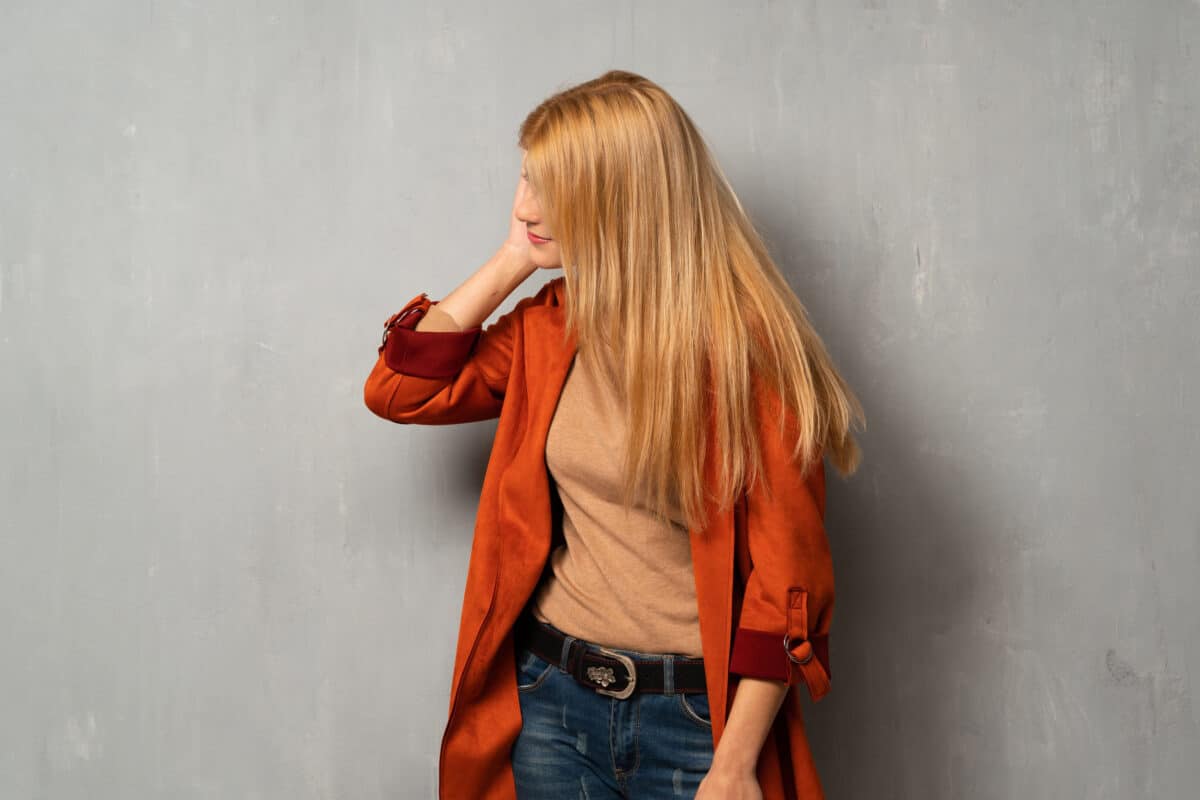
Blonde and light brown hair are more susceptible to photobleaching because they contain less melanin compared to darker hair colors. Melanin acts as a natural sunscreen, providing some protection against UV radiation. However, regardless of hair color, all hair types can experience photobleaching with sufficient sun exposure.

Interestingly, hair photobleaching does not damage the hair’s protein structure significantly. Unlike chemical bleaching, which can weaken hair, photobleaching primarily affects the melanin, leaving the hair’s physical integrity largely intact. This natural lightening process is a unique intersection of environmental influence and biological response, showcasing the dynamic relationship between nature and human physiology.

How Long Does Hair Photobleaching Take?

The sun’s rays can work wonders on hair color, lightening it over time. The duration of hair photobleaching depends on several factors, including the intensity of sun exposure, hair type, and color. On average, noticeable lightening can occur after consistent sun exposure over a few weeks. Ultraviolet (UV) radiation, particularly UVB rays, breaks down melanin, the pigment responsible for hair color. This degradation process leads to the gradual lightening of hair.

Blonde, red and light brown hair, which contain less melanin, may show signs of photobleaching more quickly than darker hair. Individuals with darker hair, which has more melanin, might require longer periods of sun exposure to see similar effects. For instance, someone with blonde hair might notice a significant change after just a few weeks of daily sun exposure, while someone with dark brown or black hair may need a month or more to see a similar effect.

Additionally, factors like time spent outdoors, hair care routines, and protective measures (such as wearing hats) can alter the timeline. Overall, while the rate of photobleaching varies, the combination of consistent sun exposure and individual hair characteristics determines how quickly the transformation occurs.

Is Hair Photobleaching Genetic?

MC1R (Melanocortin 1 Receptor)

This gene plays a significant role in determining hair color by regulating the type and amount of melanin produced in hair follicles. Variants of the MC1R gene are associated with red hair and lighter skin, making individuals more susceptible to photobleaching due to lower melanin levels. 

A 2015 study examined the genetic factors behind red hair, focusing on the melanocortin-1 receptor (MC1R) gene. It found that three specific variants of MC1R (rs1805007, rs1805008, and rs1805009) are strongly linked to red hair. Other variants (rs1805005, rs2228479, and rs885479) have a weaker connection but still contribute.

Using data from the UK Biobank, researchers confirmed that both strong and weak variants of MC1R affect hair color, but the weak variants alone can show a negative association with red hair when analyzed individually. This is because these loss-of-function variants do not appear together on the same gene copy.

The study also looked at other genes related to hair color but found that they did not significantly improve the prediction of red hair compared to using MC1R variants alone. The best model for predicting red hair based on MC1R variants was highly accurate, with a prediction success rate of 96%.

SLC45A2 Gene

This gene is involved in melanin synthesis and pigmentation. Genetic variants of SLC24A4 can influence hair color and its response to UV exposure. A 2008 study looked at how genes affect hair color, focusing on the SLC45A2 gene, which is important for melanin production. The researchers examined two specific genetic variations in SLC45A2, rs26722, and rs16891982, in a European population to see how they relate to hair color. The study found that both genetic variations are linked to differences in hair color. 

However, when both variations were analyzed together, only the rs16891982 variant (specifically, the L374F change) showed a strong association with hair color. This variation significantly increased the likelihood of having black hair, with the rare allele L374 raising the odds by over seven times. The study suggests that the L374F variation in the SLC45A2 gene is a key genetic marker for predicting black hair color which is less susceptible to photobleaching

Are Some People More Susceptible to Hair Photobleaching?

The sun’s impact on hair color varies from person to person, but certain individuals are more susceptible to hair photobleaching due to genetic, environmental, and hair characteristics. One of the primary factors is hair color. 

Individuals with lighter hair colors, such as blonde, red or light brown, are more prone to photobleaching because their hair contains less melanin. Melanin, the pigment responsible for hair color, provides natural protection against ultraviolet (UV) radiation. The less melanin present, the more vulnerable the hair is to UV-induced pigment breakdown.

Environmental factors, such as geographic location and lifestyle, also impact susceptibility to photobleaching. Those living in regions with intense sunlight or who spend a lot of time outdoors are more likely to experience hair photobleaching. 

Furthermore, the condition of the hair can affect its susceptibility. Hair that is already damaged or porous from chemical treatments may photobleach more quickly because it is more vulnerable to UV penetration. While anyone can experience hair photobleaching with sufficient sun exposure, genetic makeup, hair color, and environmental conditions play crucial roles in determining susceptibility.

Is Hair Photobleaching Bad for Your Health?

Hair photobleaching, the lightening of hair due to sun exposure, primarily affects the hair’s pigment, melanin, without significantly damaging the hair’s protein structure. The process is generally considered cosmetic and does not pose a direct threat to health. Unlike chemical bleaching, which can weaken hair and lead to breakage, photobleaching mainly impacts the melanin content, leaving the hair’s physical integrity mostly intact.

However, prolonged exposure to UV radiation from the sun, which causes photobleaching, can have adverse effects on the scalp and skin. UV radiation is a known risk factor for skin cancer, including melanoma, basal cell carcinoma, and squamous cell carcinoma. Therefore, while the lightening of hair itself is not harmful, the exposure required to achieve it can increase the risk of skin damage and skin cancer.

Additionally, excessive sun exposure can lead to dryness and brittleness in hair, making it more prone to breakage. It can also cause the scalp to become sunburned, leading to discomfort and potential long-term damage. Using protective measures, such as wearing hats and applying UV-protective hair products, can help mitigate these risks while still allowing for some natural lightening. It is important to balance enjoying the sun with protective strategies to maintain overall health and well-being.

Is Hair Photobleaching Damage Permanent?

Hair photobleaching is not a permanent condition. While this lightening effect can be long-lasting, it is not irreversible. New hair growth from the roots will retain its natural color, unaffected by previous sun exposure. As the photobleached hair is gradually cut away, the natural hair color will return.

However, sun exposure can lead to some degree of lasting damage to the hair shaft. Prolonged UV exposure can weaken the hair’s structure, making it more susceptible to dryness, brittleness, and split ends. While these effects can be managed with proper hair care, such as using moisturizing treatments and avoiding further UV exposure, the damaged hair itself does not repair or revert to its original state. Instead, maintaining healthy hair requires regular trimming to remove the photobleached and weakened ends.

Moreover, protecting hair from UV exposure using hats or UV-protective hair products can prevent further damage. Overall, while the color change from photobleaching is temporary, the structural damage from the sun exposure to the hair can be lasting, necessitating ongoing care and protection to maintain hair health.

How to Prevent Hair Photobleaching?

Preventing hair photobleaching involves minimizing exposure to UV radiation and using protective measures to shield the hair from the sun’s various damaging effects. Here are several scientifically-backed strategies:


  1. Wear hats or scarves: Covering the hair with a wide-brimmed hat or a scarf can provide a physical barrier against UV rays. This is one of the most effective ways to prevent direct sun exposure and reduce the risk of photobleaching.
  2. Use UV-protective hair products: Specialized hair care products, such as leave-in conditioners, sprays, and serums, often contain UV filters. These ingredients, like benzophenone and ethylhexyl methoxycinnamate, absorb or reflect UV radiation, protecting the hair from damage.
  3. Limit sun exposure: Reducing the time spent outdoors during peak sun hours, typically between 10 a.m. and 4 p.m., can significantly decrease UV exposure. Seeking shade when outside can also help minimize the risk of photobleaching.
  4. Stay hydrated and maintain a healthy diet: Proper hydration and a balanced diet rich in vitamins and minerals support overall hair health. Nutrients such as vitamins A, C, and E, along with omega-3 fatty acids, help maintain the integrity of the hair shaft, making it more resilient to environmental stressors.
  5. Regular conditioning treatments: Deep conditioning treatments and hair masks can help strengthen the hair and keep it moisturized. Healthy, well-moisturized hair is less prone to damage from UV exposure.
  6. Avoid chemical treatments: Limiting the use of harsh chemical treatments, such as bleaching, perming, and excessive heat styling, can reduce the hair’s vulnerability to UV damage. Chemically treated hair is often more porous and susceptible to photobleaching.

Implementing these preventive measures can help protect hair from the harmful effects of UV radiation, maintaining its natural color and health even during the sunniest months.

Ways to Treat Photobleached and Sun-Damaged Hair

Treating sun-damaged hair involves addressing both the color changes and any damage to the hair’s structure. Here are several scientifically supported methods to restore health and vibrancy to photobleached hair:

Hydrating and Moisturizing Treatments

Sun exposure often leaves hair dry and brittle. Using hydrating shampoos and conditioners can help replenish lost moisture. Ingredients like glycerin, hyaluronic acid, and aloe vera are particularly effective in attracting and retaining moisture in the hair. Deep conditioning treatments or hair masks applied once a week can provide intensive hydration and help repair damage.

Protein Treatments

UV exposure can weaken the hair’s protein structure. Protein treatments, such as those containing keratin, can help strengthen the hair shaft and reduce breakage. These treatments fill in the gaps in the hair cuticle, making the hair more resilient and smooth.

Regular Trimming

Regular trims are essential to manage and eventually eliminate damaged hair. Cutting off split ends and weakened sections prevents further breakage and promotes healthier growth. Trimming every six to eight weeks is generally recommended.

Using Leave-in Conditioners and Serums

Leave-in conditioners and serums can provide continuous protection and moisture throughout the day. Look for products with UV filters, silicones, and natural oils like argan or coconut oil, which can protect and nourish the hair while reducing frizz and improving shine.

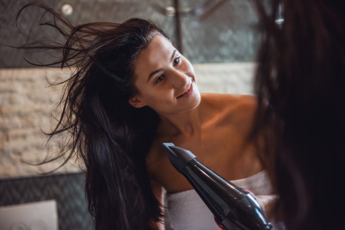
Avoiding Heat Styling

Limiting the use of heat-styling tools such as blow dryers, flat irons, and curling irons can prevent additional damage to already weakened hair. If heat styling is necessary, using a heat protectant spray can minimize damage by creating a barrier between the hair and the heat.

Color Correction

If the color change from photobleaching is undesirable, professional color correction can help restore your natural hue or achieve a new desired shade. A professional colorist can assess the extent of the bleaching and apply appropriate treatments to balance and even out the hair color.

Scalp Care

A healthy scalp is essential for healthy hair growth. Using gentle, sulfate-free shampoos and regularly massaging the scalp can improve blood circulation and promote healthier hair follicles. Products containing ingredients like tea tree oil, peppermint, and salicylic acid can help maintain a clean and healthy scalp environment.

Protective Hairstyles

Wearing protective hairstyles, such as braids, buns, or updos, can minimize hair exposure to environmental stressors and reduce the risk of further damage. These styles also help manage and protect the hair from physical manipulation and friction.

Nutritional Support

A diet rich in vitamins and minerals supports hair health from within. Ensuring adequate intake of vitamins A, C, D, E, and B-complex, along with minerals like zinc and iron, can promote stronger, healthier hair. Omega-3 fatty acids, found in fish oil and flaxseed, can also enhance hair strength and shine.

By incorporating these treatments and practices into your hair care routine, you can effectively address the damage caused by photobleaching and restore your hair’s health, strength, and luster.

LifeDNA’s Skincare Report

Discover the secrets to healthier, more radiant skin with LifeDNA’s Skincare Report. This scientifically backed and comprehensive report delves into your unique genetic profile to reveal insights tailored specifically for you. By understanding how your genetics influence your skin’s needs, you can make informed decisions about the products and routines that will work best for you.

LifeDNA’s Skincare Report is just one part of an extensive suite of reports that cover every aspect of your well-being. With over 200 individual genetic trait reports available, you can explore LifeDNA’s other main reports like Nutrition Report, Sleep Report, Wellness Report, Vitamins and Supplements Report, Fitness Report, and Personality and Cognition Report. For those seeking even deeper insights, our Premium Reports, including the Age-Related Report and Detoxification Genes Report, offer advanced understanding and guidance.

Imagine having the knowledge to tailor your skincare, fitness, and nutrition plans to your genetic makeup. LifeDNA empowers you with personalized insights, helping you optimize your lifestyle and habits based on your unique genetic blueprint. Start your wellness journey today with LifeDNA and discover the benefits of a truly personalized approach to health.

Don’t wait to unlock your genetic potential. Avail of LifeDNA’s plans now and take the first step towards a healthier, happier you. Your body is unique — your wellness plan should be too.

References

  1. https://www.ncbi.nlm.nih.gov/pmc/articles/PMC6548228/
  2. https://www.nature.com/articles/jhg2008124
  3. https://www.verywellhealth.com/hair-photobleaching-8659833
  4. https://www.fda.gov/radiation-emitting-products/tanning/ultraviolet-uv-radiation
  5. https://www.23andme.com/en-int/topics/traits/hair-photobleaching/
  6. https://www.mdanderson.org/publications/focused-on-health/what-s-the-difference-between-uva-and-uvb-rays-.h15-1592991.html#:~:text=UVB%20radiation%20makes%20up%20only,other%20types%20of%20skin%20cancer.
  7. https://my.clevelandclinic.org/health/body/22615-melanin
  8. https://www.webmd.com/beauty/what-to-know-about-hair-bleach
  9. https://www.mayoclinic.org/diseases-conditions/skin-cancer/symptoms-causes/syc-20377605
  10. https://www.verywellhealth.com/hair-photobleaching-8659833#:~:text=Without%20proper%20protection%2C%20it%20can,care%20to%20offset%20extreme%20results.
  11. https://www.bumbleandbumble.ca/uv-protection-hair-products
  12. https://www.healthline.com/health/beauty-skin-care/hyaluronic-acid-for-hair
  13. https://www.healthline.com/health/what-are-the-side-effects-of-a-keratin-treatment 

Are Flat Feet Hereditary? Exploring the Genetic Connection

Flat feet condition, showing the foot arch, exploring the genetic link between heredity and flat feet.

Disclaimer: This article is for informational purposes only and is not intended to diagnose any conditions. LifeDNA does not provide diagnostic services for any conditions mentioned in this or any other article.

Flat feet, a common condition where the arches of the feet are lowered or absent, can impact your posture and overall comfort. But have you ever wondered why some people have flat feet while others don’t? The answer might be more complicated than you think. While flat feet can result from various factors, genetics play a significant role in determining whether you’ll develop this condition. 

What are Flat Fleet?

Flat feet, or pes planus, occur when the arch of the foot collapses, causing the entire sole to make contact with the ground. This condition can be observed in both children and adults, with varying degrees of severity. 

Scientifically, flat feet can result from a variety of factors, including anatomical abnormalities, ligament laxity, or muscle weakness. In children, flat feet often develop naturally as they grow, and many cases resolve by adulthood. However, when the condition persists or causes discomfort, it might be due to genetic factors, which influence the structure and function of foot ligaments and tendons.

Research indicates that flat feet can be inherited, with studies suggesting that certain genes may contribute to the development of the condition. For instance, variations in genes responsible for collagen production, which affects the strength and flexibility of connective tissues, have been linked to flat feet. 

Additionally, biomechanical factors, such as excessive pronation or abnormal gait, can exacerbate the condition. Understanding the genetic and environmental factors contributing to flat feet is crucial for developing effective treatments and management strategies.

How to Test for Flat Feet

Testing for flat feet involves several clinical and diagnostic methods to assess the structure and function of the feet. 

  • Wet Foot Test: One of the simplest and most common tests is the Wet Foot Test. This involves wetting the feet and then stepping onto a dry surface, like a piece of paper. The imprint left can reveal the arch’s height; a complete footprint with no arch visible indicates flat feet.
  • Foot Posture Index (FPI): A more clinical approach is the Foot Posture Index (FPI), a standardized tool used by podiatrists to evaluate foot alignment and posture. The FPI involves assessing various aspects of foot structure and function through physical examination.
  • X-rays: X-rays can also be utilized to provide a detailed view of the foot’s bone structure and to measure the arch height. This method helps in diagnosing the severity of flat feet and ruling out other conditions.
  • Orthotic assessment: This test may include evaluating how the foot functions during movement. This can involve analyzing gait patterns and the distribution of weight across the foot using devices like pressure mats or force plates.

Each of these methods provides valuable information to diagnose flat feet and guide appropriate treatment strategies.

Are Flat Feet Genetic?

The genetics of flat feet reveal how inherited traits can influence the structure and function of the feet. Flat feet often arise from a combination of genetic factors and environmental influences. Understanding the genetic basis of this condition provides insights into why some people develop flat feet while others do not.

Key genetic markers associated with flat feet include variations in genes responsible for collagen production, such as COL1A1 and COL1A2. Collagen is crucial for maintaining the strength and elasticity of tendons and ligaments, including those that support the arch of the foot. Alterations in these genes can weaken the connective tissues, leading to a collapsed arch.

Another important gene is the GDF5 gene, which plays a role in the development of bones and joints. Variants in GDF5 have been linked to various skeletal disorders, including abnormalities in foot arch formation. Similarly, the FOXP1 gene, involved in skeletal development, has associations with structural abnormalities in the foot.

The PITX1 gene is also relevant, as it affects limb and foot development. Mutations or variations in PITX1 can impact the formation of the arch. Additionally, the ANKRD1 gene, related to musculoskeletal development, has been linked to conditions that can influence foot structure.

While genetics play a significant role, flat feet can also be influenced by factors such as muscle weakness, injury, or obesity. Understanding these genetic links helps in managing and addressing flat feet effectively.

What are Other Causes of Flat Feet?

Flat feet, while often genetic, can arise from various other causes that affect the foot’s structure and function. Understanding these causes is crucial for effective management and treatment.

  1. Posterior Tibial Tendon Dysfunction (PTTD): This condition involves the degeneration or inflammation of the posterior tibial tendon, which supports the arch. Over time, this tendon may weaken, leading to the collapse of the foot’s arch. PTTD is a common cause of acquired flat feet and is often seen in middle-aged individuals.
  2. Trauma or Injury: Injuries to the foot, such as fractures or sprains, can damage the structures that maintain the arch. For instance, a severe injury to the bones or ligaments can disrupt normal foot mechanics and lead to flat feet.
  3. Arthritis: Various forms of arthritis, including rheumatoid arthritis, can affect the joints and ligaments in the feet. Chronic inflammation and joint damage can contribute to the development of flat feet by compromising the integrity of the arch-supporting structures.
  4. Muscle Weakness or Imbalance: Conditions that weaken or imbalance the muscles supporting the foot, such as muscular dystrophy or cerebral palsy, can lead to flat feet. The loss of muscle strength or coordination impacts the foot’s ability to maintain its arch.
  5. Congenital Conditions: Some individuals are born with structural abnormalities that predispose them to flat feet. For example, conditions such as congenital talipes equinovarus (clubfoot) can affect the normal development of the foot arch.
  6. Obesity: Excess body weight increases the stress on the feet, potentially leading to the collapse of the arch over time. The additional load can exacerbate existing issues or contribute to the development of flat feet.
  7. Diabetes: Long-term diabetes can lead to diabetic neuropathy, which affects nerve function in the feet. This condition can alter foot mechanics and contribute to the development of flat feet due to changes in sensory feedback and muscle control.
  8. Flat Foot Syndrome in Children: While many children have flat feet that resolve naturally, some may experience persistent flat feet due to developmental issues. Conditions such as flexible flatfoot, where the arch appears normal when not bearing weight, can be observed in young children.

These diverse causes highlight the importance of a comprehensive evaluation when diagnosing and managing flat feet, ensuring that treatment strategies address the specific underlying factors.

What are the Types of Flat Fleet?

Flat feet come in several types, each with distinct characteristics and underlying causes. Understanding these types helps diagnose and manage the condition effectively.

  • Flexible Flatfoot: This is the most common type and is characterized by a visible arch when the foot is not bearing weight but appears flat when standing. It is often seen in children and tends to resolve with age as the foot structure matures. Flexible flatfoot usually does not cause pain or significant functional issues but can be monitored for any potential complications.
  • Rigid Flatfoot: Unlike flexible flatfoot, rigid flatfoot presents with a consistently flat arch regardless of the foot’s position. This type often results from structural abnormalities or deformities, such as a fused bone or a severely collapsed arch. Rigid flatfoot can cause discomfort and may require medical intervention or surgery to correct.
  • Congenital Flatfoot: This type is present at birth and results from developmental abnormalities in the foot’s structure. Congenital flatfoot can be due to conditions like clubfoot (congenital talipes equinovarus) or abnormal bone development. Early diagnosis and treatment are essential to manage and potentially correct the deformity.
  • Acquired Flatfoot: Acquired flatfoot develops later in life due to external factors such as injury, arthritis, or tendon dysfunction. Posterior Tibial Tendon Dysfunction (PTTD) is a common cause of acquired flatfoot, where the tendon supporting the arch becomes damaged or weakened, leading to the collapse of the arch.
  • Adult-Acquired Flatfoot: This type specifically refers to flatfoot that develops in adults, often due to conditions like PTTD or age-related degeneration. It can be progressive and cause pain, requiring a tailored treatment approach to address the underlying causes and symptoms.
  • Flexible Flatfoot with an Ankle Foot Orthosis (AFO): This type is a variant of flexible flatfoot where the use of an orthotic device, such as an AFO, helps maintain the arch and alleviate symptoms. It is used in cases where additional support is needed for comfort or functional purposes.

Each type of flatfoot presents unique challenges and treatment needs, making accurate diagnosis crucial for effective management.

Are Flat Feet Dangerous to Your Health?

Flat feet, in themselves, are not necessarily dangerous to health, but they can lead to various issues if left untreated. When the arches of the feet collapse, it can affect the alignment and biomechanics of the entire lower limb. This misalignment may cause or exacerbate conditions such as plantar fasciitis, where the tissue connecting the heel to the toes becomes inflamed, leading to heel pain.

Flat feet can also contribute to shin splints and knee pain due to altered gait mechanics. The lack of proper arch support can place excessive strain on the knees and hips, potentially leading to joint pain and discomfort. In severe cases, ankle instability and lower back pain may also arise from the improper distribution of forces during movement.

For individuals with pre-existing conditions like arthritis or diabetes, flat feet can complicate the management of these conditions by adding additional stress on the lower limbs. Although flat feet are often manageable with appropriate interventions such as orthotics or physical therapy, neglecting to address the associated symptoms may lead to chronic pain and functional limitations. Regular evaluation by a healthcare professional is recommended to prevent complications and ensure proper management.

Are Flat Feet Curable?

Flat feet are generally not considered curable in the traditional sense, but their symptoms can often be managed effectively. Treatment typically focuses on alleviating discomfort, improving function, and preventing complications rather than completely reversing the condition.

For children with flexible flatfoot, the condition often improves naturally as they grow, and many do not require intervention. However, if symptoms persist or cause pain, treatment options like orthotic insoles, physical therapy, and exercises to strengthen foot and leg muscles can help manage the condition.

In cases of rigid flatfoot or severe flatfoot resulting from conditions such as posterior tibial tendon dysfunction, treatment may involve more comprehensive approaches. Orthotic devices can provide support and improve foot mechanics, while physical therapy can address muscle imbalances and improve flexibility. In some situations, surgical intervention may be considered to correct structural abnormalities or repair damaged tendons.

For adult-acquired flatfoot, addressing the underlying cause—such as arthritis or tendon dysfunction—is crucial. While complete reversal of the condition may not be possible, appropriate management can significantly reduce pain and improve quality of life. Regular follow-up with a healthcare professional helps ensure the effectiveness of the chosen treatment strategy.

Symptoms of Flat Feet

Flat feet can present with a variety of symptoms, which can vary depending on the severity of the condition and any underlying causes. Common symptoms may include:

  • Foot Pain: Individuals with flat feet often experience discomfort in the arch or heel area. This painful condition, known as plantar fasciitis, results from the inflammation of the tissue connecting the heel to the toes.
  • Arch Pain: The collapse of the arch can lead to localized pain and tenderness along the inner part of the foot where the arch is absent or weakened.
  • Swelling: Swelling may occur in the feet, particularly around the ankle, due to the added strain and altered mechanics of foot movement.
  • Overpronation: This refers to the excessive inward rolling of the foot during walking or running, which can lead to further strain on the foot and lower limb joints.
  • Knee and Hip Pain: The misalignment caused by flat feet can affect the knees and hips, leading to pain and discomfort in these areas due to altered gait and increased stress.
  • Lower Back Pain: The altered biomechanics from flat feet can also impact the lower back, causing discomfort and pain due to compensatory changes in posture.
  • Difficulty Walking or Standing: Individuals may find it challenging to walk long distances or stand for extended periods due to fatigue and discomfort.
  • Foot Fatigue: People with flat feet often experience increased fatigue in the feet and legs, especially after physical activities.

Recognizing these symptoms and seeking appropriate treatment can help manage and alleviate the discomfort associated with flat feet.

Ways to Manage Flat Feet

Managing flat feet involves a range of strategies aimed at alleviating symptoms and improving foot function. The choice of management techniques depends on the severity of the condition and any associated symptoms.

Orthotic Devices

Custom or over-the-counter orthotic insoles provide additional arch support and help correct foot alignment. They can reduce pain, improve gait, and redistribute pressure more evenly across the foot. Research shows that orthotics can be effective in managing symptoms of flat feet, particularly in reducing plantar fasciitis and foot fatigue.

Footwear

Wearing supportive shoes is crucial for managing flat feet. Shoes with good arch support, cushioning, and stability features help maintain proper foot alignment and reduce strain. Avoiding shoes with inadequate support, such as flip-flops or high heels, can prevent exacerbation of symptoms.

Physical Therapy

Engaging in physical therapy can strengthen the muscles supporting the foot and improve flexibility. Exercises often include stretching the Achilles tendon and calf muscles, strengthening the posterior tibial tendon, and improving overall foot mechanics. Clinical studies have demonstrated that targeted physical therapy can alleviate symptoms and improve function in individuals with flat feet.

Foot Exercises

Specific exercises, such as toe curls, arch lifts, and foot rolling, can help strengthen foot muscles and support structures. Regularly performing these exercises can improve the overall function of the foot and reduce discomfort.

Weight Management

Maintaining a healthy weight can reduce the stress and pressure on the feet. Excess body weight can exacerbate the symptoms of flat feet by increasing the load on the arches and altering foot mechanics.

Pain Management

Over-the-counter pain relievers, such as nonsteroidal anti-inflammatory drugs (NSAIDs), can be used to manage pain and inflammation associated with flat feet. For chronic pain, a healthcare provider might recommend other treatments or medications.

Physical Modifications

Using cushioned insoles or arch supports in everyday footwear can provide immediate relief and improve comfort. Adjustments in activity levels and the use of supportive devices during physical activities can also help manage symptoms.

Surgical Intervention

In severe cases where conservative measures are insufficient, surgical options might be considered. Surgical procedures can involve correcting structural abnormalities, repairing damaged tendons, or reconstructing the arch. Surgery is typically reserved for cases where flat feet cause significant pain or functional limitations that do not respond to other treatments.

These management strategies can help individuals with flat feet lead a more comfortable and active life. Regular consultation with healthcare professionals ensures that the chosen approach effectively addresses the symptoms and underlying causes.

Why Choose LifeDNA

With LifeDNA, you gain access to a range of detailed reports, including Nutrition Report, Wellness Report, Sleep Report, Vitamins and Supplements Report, Fitness Report, Personality and Cognition Report, and Skincare, along with premium options like the Age-Related Report, Methylation Genes Report, and Detoxification Genes Report. These reports delve into nearly 200 specific traits, guiding you on how to optimize your lifestyle based on your genetic makeup.

Imagine understanding exactly what vitamins your body needs, how to tailor your fitness routine for maximum effectiveness, or discovering personalized nutrition strategies. LifeDNA’s DNA Trait Reports help you make informed decisions that align with your genetic profile, setting you on the path to a healthier, more balanced life.

Start your journey toward personalized wellness today. Explore LifeDNA’s plans and take control of your health with insights designed just for you. Embrace the power of genetic information and make choices that are right for your unique body.

References

  1. https://my.clevelandclinic.org/health/diseases/17005-flat-feet
  2. https://www.nhs.uk/conditions/flat-feet/#:~:text=To%20see%20if%20you%20have,be%20flat%20on%20the%20ground.&text=You%20do%20not%20have%20flat,this%20is%20called%20the%20arch).
  3. https://www.physio-pedia.com/Foot_Posture_Index_(FP1-6)
  4. https://uddingstonphysiotherapy.co.uk/orthotic-assessment/#:~:text=An%20orthotist%20assesses%20joints%2C%20ligaments,help%20to%20strengthen%20soft%20tissues.
  5. https://www.sciencedirect.com/topics/nursing-and-health-professions/flatfoot
  6. https://pubmed.ncbi.nlm.nih.gov/16892395/
  7. https://pubmed.ncbi.nlm.nih.gov/34131944/
  8. https://www.sciencedirect.com/topics/biochemistry-genetics-and-molecular-biology/pitx1
  9. https://www.ncbi.nlm.nih.gov/gene/27063
  10. https://my.clevelandclinic.org/health/diseases/22057-posterior-tibial-tendon-dysfunction
  11. https://www.foothealthfacts.org/conditions/osteoarthritis-of-the-foot-and-ankle#:~:text=A%20flat%20foot%20causes%20less,an%20increased%20risk%20of%20arthritis.
  12. https://www.azfootdocs.com/blog/the-link-between-obesity-and-flat-feet#:~:text=Obesity%20is%20another%20factor%20that,which%20can%20cause%20flat%20feet.
  13. https://dergipark.org.tr/en/pub/turkjkin/issue/81884/1358860#:~:text=Diabetic%20patients%20are%20more%20prone,the%20course%20of%20their%20disease.
  14. https://www.chop.edu/conditions-diseases/flat-feet-in-children#:~:text=Flat%20feet%20%E2%80%94%20or%20pes%20planus,feet%20haven’t%20yet%20developed.
  15. https://www.medparkhospital.com/en-US/disease-and-treatment/flat-feet
  16. https://www.mdpi.com/1996-1944/14/18/5297
  17. https://www.advancedfootdocs.com/blog/why-flat-feet-are-bad-for-your-health/#:~:text=Your%20body’s%20balance%20begins%20in,ankles%2C%20knees%2C%20or%20hips.
  18. https://www.bauerfeind.com.au/blogs/news/can-flat-feet-be-cured#:~:text=Thankfully%2C%20in%20many%20cases%2C%20flat,caused%20by%20lifestyle%20or%20injury).
  19. https://www.nyp.org/orthopedics/columbia-orthopedics/flat-feet
  20. https://study.com/academy/lesson/orthotic-devices-types-functions-examples.html#:~:text=Orthotic%20devices%2C%20also%20called%20orthoses,immobilization%20and%20alignment
  21. https://www.healthline.com/health/flat-feet-exercises
  22. https://www.medicalnewstoday.com/articles/flat-foot-reconstruction-surgery#:~:text=Flat%20foot%20surgery%20aims%20to,foot%20or%20unusually%20low%20arches. 

How Genetics Influence Platelet Count

Zoomed-in view of blood cells, illustrating platelet count and the genetic factors influencing platelet count production and regulation

Disclaimer: This article is for informational purposes only and is not intended for diagnostic use. LifeDNA does not provide diagnostic reports on any traits discussed. Genetics is just one piece of the puzzle; please consult a healthcare professional for comprehensive guidance on any health condition.

Platelets, those tiny cell fragments in your blood, play a crucial role in clotting and healing wounds. When your platelet count is too low or too high, it can signal various health issues. But what determines your platelet count? Could your genetics affect something this fundamental?  

What is a Platelet?

Blood is more than just a red liquid; it’s a complex system full of fascinating components. Among these are platelets, tiny but mighty players in the body’s defense mechanism against bleeding. Platelets, also known as thrombocytes, are small, colorless cell fragments in the blood that are crucial for blood clotting and wound healing. Unlike red and white blood cells, human platelets do not have a nucleus. They are produced in the bone marrow from large cells called megakaryocytes, which release thousands of platelets into the bloodstream. 

Platelets circulate in the blood and are always on standby to respond to blood vessel injuries. When a blood vessel is damaged, platelets rush to the site and adhere to the vessel’s lining. They release chemicals that activate other platelets and initiate the formation of a platelet plug, a temporary seal to prevent further bleeding. 

Additionally, platelets release substances that interact with proteins in the blood plasma to form a more stable clot, a fibrin clot. Understanding platelet function and count is essential in diagnosing and managing many hematologic and cardiovascular diseases.

What is a Platelet Count?

Platelet counts reveal critical information about a person’s blood health and potential underlying conditions. A platelet count measures the number of platelets, or thrombocytes, in a microliter of blood. These tiny, disc-shaped cells are vital for blood clotting and wound repair. 

Blood samples for platelet counts are often analyzed using automated hematology analyzers. These machines provide quick and accurate results, which are essential for diagnosing and monitoring various medical conditions.

Monitoring platelet counts helps healthcare providers manage health risks and tailor treatments accordingly. Regular platelet count tests are crucial for patients with chronic illnesses, those undergoing certain treatments, or individuals with symptoms indicative of platelet disorders.

How to Test for Platelet Count

Testing for platelet count is a straightforward yet crucial procedure in medical diagnostics. The process usually begins with a complete blood count (CBC) test, which includes a platelet count as one of its components. A healthcare professional collects a blood sample, typically from a vein in the arm, using a sterile needle. The blood sample is then sent to a laboratory for analysis. 

In the lab, automated hematology analyzers are commonly used to count platelets. These machines work by measuring the electrical impedance or light scattering properties of the blood cells. The results provide a platelet count, expressed as the number of platelets per microliter of blood. Normal platelet counts range from 150,000 to 450,000 platelets per microliter. 

Platelet counts outside this range can indicate various medical conditions. Low platelet counts, or thrombocytopenia, can be caused by bone marrow disorders, autoimmune diseases, certain infections, or medications. Symptoms of low platelet counts include easy bruising, prolonged bleeding, and spontaneous bleeding from the gums or nose. 

 Conversely, high platelet counts, or thrombocytosis, can result from bone marrow disorders, inflammation, infection, or cancer. Elevated platelet levels increase the risk of abnormal blood clots, which can lead to strokes or heart attacks. Regular monitoring of platelet counts helps in the diagnosis and management of these conditions, guiding appropriate medical intervention.

Is Platelet Count Genetic?

JAK2 Gene

The Janus Kinase 2 gene (JAK2) provides instructions for making a protein that regulates blood cell production in the bone marrow. Mutations in JAK2 can lead to myeloproliferative neoplasms (MPNs), where the bone marrow produces excessive blood cells. Common MPNs linked to JAK2 mutations include:

  • Polycythemia Vera (PV): Excessive red blood cells.
  • Essential Thrombocythemia (ET): Overproduction of platelets.
  • Primary Myelofibrosis (PMF): Excess platelets and scar tissue in the bone marrow.


The JAK2 V617F mutation is the most studied, causing the JAK2 protein to be constantly active, leading to uncontrolled blood cell production. Other mutations in JAK2 can also contribute to these disorders, with tests available to detect various JAK2 mutations, including those in exon 12.

CALR Gene

In essential thrombocythemia (ET), patients with CALR mutations exhibit a lower risk of thrombosis compared to those with JAK2V617F mutations. Research reveals that CALR-mutated platelets are less activated and adhere less to fibrinogen than control and JAK2-mutated platelets. Specifically, fewer CALR-mutated platelets fully spread on fibrinogen.


CALR mutations are linked to a more dispersed localization of the CALR protein and a unique interaction with TpoR, which affects platelet function. Despite higher platelet counts and increased immature platelets and platelet-leukocyte aggregates in ET patients overall, CALR mutations result in less activated platelets, which may explain the reduced thrombotic risk in these patients.

What are Other Factors AffectingPlatelet Count Levels?

Platelet counts can be influenced by various factors beyond genetic predispositions. Understanding these factors provides a more comprehensive view of what affects platelet levels and how they can impact overall health.

Medical Conditions

  • Bone Marrow Disorders: Conditions such as leukemia and myelodysplastic syndromes can significantly reduce platelet production. Conversely, myeloproliferative disorders, where the bone marrow produces too many blood cells, can lead to elevated platelet counts.
  • Autoimmune Diseases: Diseases like lupus and rheumatoid arthritis can cause the immune system to attack platelets, leading to thrombocytopenia.
  • Infections: Viral infections, particularly those caused by viruses like dengue, HIV, and hepatitis, can reduce platelet numbers either directly or through an immune-mediated process.
  • Liver Diseases: Conditions such as cirrhosis can cause splenic sequestration of platelets, reducing their numbers in circulation.

Medications

  • Chemotherapy Drugs: These can suppress bone marrow function, leading to reduced platelet production.
  • Heparin: This anticoagulant can cause heparin-induced thrombocytopenia (HIT), a condition where the immune system forms antibodies against platelets.
  • Antibiotics: Some antibiotics, like vancomycin, can cause thrombocytopenia through immune-mediated mechanisms.
  • Non-Steroidal Anti-Inflammatory Drugs (NSAIDs): These can impair platelet function, although they do not typically reduce significantly platelet numbers.

Nutritional Deficiencies

  • Vitamin B12 and Folate Deficiency: These are essential for DNA synthesis in bone marrow cells, including megakaryocytes, which produce platelets. Deficiencies can result in reduced platelet production.
  • Iron Deficiency: Severe iron deficiency can also impact platelet production and function.

Lifestyle Factors

  • Alcohol Consumption: Excessive alcohol intake can lead to bone marrow suppression and reduced platelet production.
  • Smoking: Smoking has been linked to increased platelet activation and higher platelet counts, which may elevate the risk of clot formation.

Surgery and Trauma

  • Major Surgeries: These can lead to significant blood loss and consumption of platelets in clot formation, temporarily lowering platelet counts.
  • Trauma: Severe trauma can cause similar reductions in platelet numbers due to bleeding and platelet consumption.

Pregnancy

  • Gestational Thrombocytopenia: A mild drop in platelet count is common in pregnancy due to increased plasma volume, which dilutes the blood. Rarely, more severe thrombocytopenia can occur due to conditions like preeclampsia or HELLP syndrome.

Understanding these diverse factors is crucial for diagnosing and managing conditions that affect platelet levels, ensuring that patients receive appropriate care based on the underlying causes.

What Do High and Low Platelet Counts Mean?

Platelets are vital for blood clotting, and their counts can reveal important health information. What do high and low platelet counts mean for your body?

Thrombocytosis

High platelet counts, known as thrombocytosis, can be caused by various conditions. Primary thrombocytosis is due to bone marrow disorders like essential thrombocythemia or polycythemia vera, where the bone marrow produces too many platelets. 

Secondary thrombocytosis is a reactive condition, often caused by inflammation, infection, cancer, or recent surgery. Elevated platelet levels can increase the risk of clot formation, leading to complications such as deep vein thrombosis, pulmonary embolism, heart attacks, or strokes. 

Symptoms of high platelet counts may include headaches, dizziness, chest pain, and tingling in the hands and feet. In some cases, no symptoms are present, and thrombocytosis is detected incidentally during routine blood tests.

Thrombocytopenia

Low platelet counts, or thrombocytopenia, can result from several factors. Bone marrow disorders, such as leukemia or aplastic anemia, can reduce platelet production. Autoimmune diseases like immune thrombocytopenic purpura (ITP) cause the body to destroy its platelets. 

Certain medications, including chemotherapy drugs and heparin, can also lead to thrombocytopenia. Additionally, viral infections such as dengue, hepatitis, and HIV can reduce platelet counts. 

Symptoms of low platelet counts include easy bruising, prolonged bleeding from cuts, spontaneous bleeding from the gums or nose, and the appearance of petechiae, which are small red or purple spots on the skin caused by bleeding.

Managing high or low platelet counts involves addressing the underlying cause. For thrombocytosis, treatment may include medications to reduce platelet production or blood thinners to prevent clots. 

Thrombocytopenia management might involve corticosteroids to suppress the immune system, medications to increase platelet production, or transfusions in severe cases. Regular monitoring and follow-up with healthcare providers are essential for managing platelet count abnormalities and minimizing the risk of complications. Understanding what high and low platelet counts mean can help individuals take proactive steps to maintain their health.

Ways to Maintain Normal Platelet Count

Maintaining a normal platelet count is essential for overall health, as platelets play a critical role in blood clotting and wound healing. Here are some scientifically-backed ways to maintain healthy platelet levels:

Balanced Diet

A nutritious diet rich in vitamins and minerals is crucial for maintaining normal platelet counts.

  • Vitamin B12: Found in animal products like meat, eggs, and dairy, vitamin B12 is essential for platelet production. Deficiency can lead to low platelet counts.
  • Folate (Vitamin B9): Crucial for DNA synthesis in bone marrow cells, folate is abundant in leafy greens, citrus fruits, and beans.
  • Iron: Necessary for hemoglobin production, iron can be obtained from red meat, poultry, fish, lentils, and fortified cereals. Severe iron deficiency can impact platelet production.
  • Vitamin C: Enhances iron absorption and supports the immune system. Sources include citrus fruits, strawberries, bell peppers, and broccoli.
  • Vitamin K: Plays a role in blood clotting. It is found in leafy greens, such as spinach, kale, and broccoli.

Avoid Alcohol and Smoking

Excessive alcohol consumption can suppress bone marrow function, reducing platelet production. Smoking is associated with increased platelet activation and can elevate the risk of clot formation.

Stay Hydrated

Adequate hydration supports overall cellular function, including platelet production. Aim to drink at least 8 glasses of water daily.

Regular Exercise

Engaging in moderate physical activity helps improve circulation and overall cardiovascular health, indirectly supporting healthy platelet levels. However, avoid excessive or high-risk activities that might lead to injury and bleeding.

Manage Stress

Chronic stress can negatively impact the immune system and potentially affect platelet levels. Techniques such as meditation, yoga, and deep-breathing exercises can help manage stress effectively.

Avoid Toxins and Chemicals

Exposure to certain chemicals, including pesticides and industrial solvents, can harm bone marrow function and reduce platelet production. Use protective gear and follow safety guidelines if you work in environments with such exposures.

Regular Medical Check-ups

Routine blood tests can monitor platelet levels and help detect any abnormalities early. Regular check-ups are especially important for individuals with a history of platelet disorders or other health conditions affecting platelet counts.

Medication Management

Some medications can affect platelet counts. Always inform your healthcare provider about all the medications and supplements you are taking. Avoid over-the-counter NSAIDs like aspirin and ibuprofen, which can impair platelet function, unless prescribed by a doctor.

Treat Underlying Conditions

Addressing health issues that can affect platelet counts is crucial. For instance, managing autoimmune diseases, liver conditions, and infections can help maintain normal platelet levels.

Herbal Supplements and Natural Remedies

Certain natural substances are believed to support healthy platelet counts, though more research is needed.

  • Papaya Leaf Extract: Some studies suggest it may increase platelet counts, especially in dengue fever cases with thrombocytopenia.
  • Aloe Vera: Known for its anti-inflammatory properties, aloe vera may support overall immune health.
  • Wheatgrass Juice: Rich in chlorophyll, it may help increase platelet production.

Avoid Unnecessary Blood Loss

Minimize the risk of injuries and avoid activities that may lead to cuts or bruises, especially if you already have a lower platelet count.

Maintaining normal platelet counts involves a combination of healthy lifestyle choices, proper nutrition, and regular medical care. By incorporating these strategies, individuals can support their body’s natural ability to produce and maintain adequate platelet levels, ensuring optimal blood clotting and overall health.

LifeDNA’s Nutritional Report

Discover the power of personalized wellness with LifeDNA’s Nutrition Report. This scientifically backed and comprehensive report delves into your unique genetic profile to help you understand your body’s specific needs. By analyzing more than 200 genetic traits, LifeDNA provides insights across seven main categories: Nutrition Report, Wellness Report, Sleep Report, Vitamins and Supplements Report, Fitness Report, Personality and Cognition Report, and Skincare. Premium insights like the Age-Related Report, Methylation Genes Report and Detoxification Genes Report are also available.

With LifeDNA’s Genetics Reports, you can uncover how your genetics influence your nutritional requirements, fitness potential, and overall well-being. Imagine knowing which vitamins and supplements your body needs the most, optimizing your sleep patterns based on genetic tendencies, or understanding your skin’s unique needs for healthier, more radiant skin. 

Start your wellness journey with LifeDNA today. Empower yourself with the knowledge to make informed lifestyle choices and achieve your health goals. Avail of LifeDNA’s plans now and unlock the full potential of your genetic insights. Take the first step toward a healthier, more personalized approach to your well-being.

Experience the benefits of knowing your body like never before. Begin your journey with LifeDNA’s Nutrition Report and embrace a future of optimized health and wellness tailored just for you.

References

  1. https://www.ironwoodcrc.com/boost-your-platelets-with-nutrition/#:~:text=Clinical%20research%20reported%20that%20Aloe,platelet%20count%20in%20your%20blood.
  2. https://www.mountsinai.org/health-library/tests/platelet-count#:~:text=A%20platelet%20count%20is%20a,red%20or%20white%20blood%20cells.
  3. https://my.clevelandclinic.org/health/diagnostics/21782-platelet-count
  4. https://www.hopkinsmedicine.org/health/conditions-and-diseases/what-are-platelets-and-why-are-they-important
  5. https://www.sciencedirect.com/topics/nursing-and-health-professions/myeloproliferative-disorder#:~:text=MYELOPROLIFERATIVE%20DISORDERS,-The%20myeloproliferative%20disorders&text=As%20many%20as%2015%25%20of,can%20occur%20in%20essential%20thrombocythemia.
  6. https://cancer.ca/en/cancer-information/cancer-types/leukemia/what-is-leukemia/myelodysplastic-syndromes#:~:text=Many%20people%20with%20an%20MDS,fever
  7. https://www.ncbi.nlm.nih.gov/gene/4352#gene-expression
  8. https://www.testing.com/tests/jak2-mutation/#:~:text=The%20JAK2%20mutation%20test%20is,thrombocythemia%20(ET)%2C%20or%20primary
  9. https://onlinelibrary.wiley.com/doi/full/10.1002/ajh.25713
  10. https://www.healthline.com/health/lupus/lupus-low-platelets#:~:text=With%20lupus%2C%20you%20may%20have,lower%20than%20150%2C000%20per%20microliter%20.
  11. https://www.healthline.com/health/cancer/minimum-platelet-count-for-chemotherapy#:~:text=Chemicals%20in%20chemotherapy%20drugs%20can,normal%20within%204%E2%80%936%20days.
  12. https://ashpublications.org/hematology/article/2009/1/225/19808/Heparin-induced-thrombocytopenia-when-a-low
  13. https://www.ncbi.nlm.nih.gov/pmc/articles/PMC5394486/
  14. https://www.mayoclinic.org/diseases-conditions/thrombocytosis/symptoms-causes/syc-20378315#:~:text=Overview,condition%2C%20such%20as%20an%20infection.
  15. https://www.mayoclinic.org/diseases-conditions/thrombocytopenia/symptoms-causes/syc-20378293
  16. https://www.news-medical.net/health/Papaya-Leaf-Extract-and-Platelet-Count.aspx
  17. https://www.ironwoodcrc.com/boost-your-platelets-with-nutrition/#:~:text=Clinical%20research%20reported%20that%20Aloe,platelet%20count%20in%20your%20blood.Â