Is There a Genetic Cause Behind Cataracts?
![]() Catherine
on
May 8, 2025
Catherine
on
May 8, 2025

Disclaimer: This article is for informational purposes only and is not intended to diagnose any conditions. LifeDNA does not provide diagnostic services for any conditions mentioned in this or any other article.
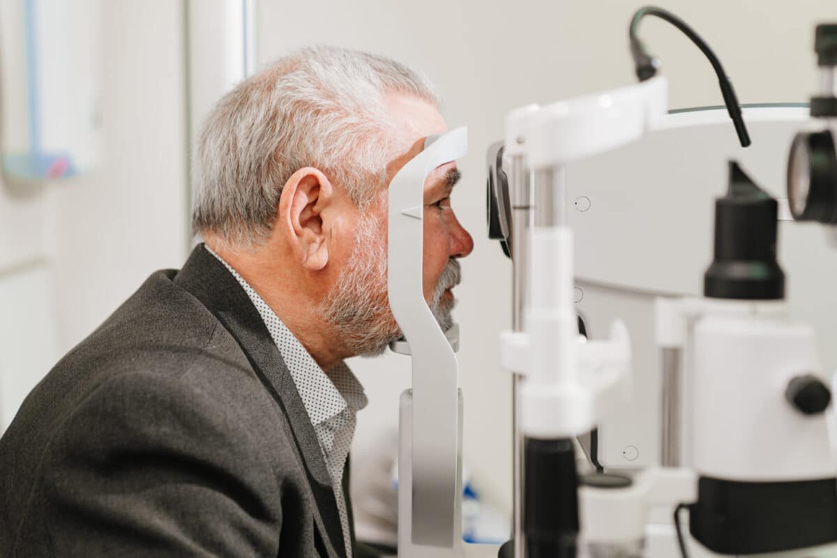
Having cataracts may feel like looking through a foggy window as your vision becomes cloudy, colors look dull, and it gets harder to read or see clearly at night. While cataracts often happen as people get older, not everyone experiences them the same way. Some people start noticing changes earlier in life, especially if family members also had cataracts. This raises an important question. Can cataracts be passed down through genes?
Aging is the most common cause of cataracts, but genetics may also play a role. Understanding this connection matters because it may help you stay ahead of vision problems, motivate you to protect your eyes from sunlight and manage other health conditions, so that you can keep your eyesight as good as possible into an advanced age.
You may want to read: Genetics of Color Blindness
What Are Cataracts?
A cataract occurs when the normally clear lens of the eye becomes cloudy. This clouding blocks light from passing through the lens properly, which results in vision problems. Many people describe it like looking through a foggy window. Cataracts are very common as you get older. In fact, more than half of all Americans age 80 or older either have cataracts or have had surgery to get rid of cataracts.
Congenital cataracts are rare as they happen only in about 72 out of every 100,000 children. About 8–25% of these cases are inherited, meaning it is possible that this condition may be passed down from parents to children. Congenital cataracts may appear on their own or as part of a syndrome involving other health conditions.
A cataract may develop slowly and may not affect vision at first. But over time, it may interfere with daily activities such as reading, driving, or recognizing faces.
What Are the Types of Cataracts?
A cataract may develop in different parts of the lens, and each type can affect vision in a unique way. Understanding the types of cataracts can help explain how symptoms may vary from person to person.
Nuclear Cataracts: These form in the center of the lens and are the most common type associated with aging. In the early stages, they might actually improve your reading vision, a phenomenon known as “second sight.” However, over time, the lens becomes yellow or brown, making distance vision blurry and colors harder to distinguish.
Cortical Cataracts: Cortical cataracts begin as white, wedge-shaped streaks around the outer edge of the lens. As they progress, the streaks move toward the center, disrupting how light passes through the lens. This type often causes problems with glare and contrast, especially in bright light.
Posterior Subcapsular Cataracts: These form at the back of the lens, right in the path of incoming light. They tend to develop faster than other types and can interfere with reading, cause glare or halos around lights, and make vision in bright environments difficult. They’re often seen in people taking steroids or those with diabetes.
Congenital Cataracts: These are present at birth or develop in early childhood. They may be inherited or result from infections, trauma, or health conditions during pregnancy. Not all congenital cataracts affect vision, but if they do, early treatment is needed to prevent long-term vision problems.
What Causes Cataracts?
Most cataracts develop as a result of aging. As we grow older, the proteins and fibers in the lens of the eye begin to break down, causing cloudiness. But aging isn’t the only factor. Other causes of cataracts include:
- Eye Injuries: Trauma to the eye, whether from an accident, blunt force, or sharp object, may damage the lens and lead to cataract formation. Sometimes the cataract may appear soon after the injury, or it may develop years later as a result of internal damage or inflammation.
- Previous Eye Surgery: Surgeries done to treat other eye problems, such as retinal disorders may increase the risk of developing cataracts later on. The procedure may disrupt the natural structure of the eye or trigger changes in the lens that lead to clouding.
- Diabetes: People with diabetes have a higher risk of cataracts due to prolonged high blood sugar levels, which may change the structure of the lens. These changes make the lens more likely to become cloudy over time, especially if blood sugar isn’t well controlled.
- Long-Term Use of Corticosteroid Medications: Extended use of corticosteroids, whether taken orally, through inhalers, or as eye drops has been linked to cataract development. These medications can interfere with the normal function of the lens and speed up clouding, especially with high doses or long durations.
- Prolonged Exposure to Ultraviolet (UV) Light: Spending long hours in the sun without eye protection may increase cataract risk. UV rays may cause damage to the proteins in the lens, leading to changes that result in cloudiness and impaired vision over time.
- Smoking and Heavy Alcohol Consumption: Smoking introduces harmful chemicals into the body that may damage the eye’s lens, while excessive alcohol intake may deplete antioxidants that help maintain eye health.
You may want to read: How to Improve Eye Health Naturally With Vitamin A
Genetics on Cataracts
Studies show that certain genetic variants may influence whether a person develops cataracts early in life or later on with age. In some cases, the same gene is involved in both thecongenital (inherited) and age-related cataracts, depending on how serious the gene changes are. For example, changes in the GALK1 gene, which helps process sugars in the body, may cause cataracts in babies when both copies of the gene from the parents are affected. But even people who only carry one altered copy—like a parent of an affected child, may have a higher risk of developing cataracts as they age. One variation of this gene, called the “Osaka variant,” is linked to a higher rate of cataracts in Japanese adults.
Other genes also play a role. The SLC16A12 gene, which helps transport nutrients and waste, has mutations that may cause childhood cataracts along with other issues like sugar in the urine and small eye size. Milder variants in the same gene have been found in adults with age-related cataracts. Similarly, EPHA2, a gene involved in lens structure and development, is connected to both early-onset and age-related cataracts. One variant in this gene reduces its activity and may increase a person’s risk over time.
Another well-studied gene is CRYAA, which produces α-crystallin which is a a protein that helps keep the lens clear by preventing other proteins from clumping. A specific variation in this gene lowers its protective effect, making it harder for the lens to fight off damage, especially in older adults. This gene, along with others like SOX2, TMPRSS5, and BICRA, has been linked to nuclear cataracts, which affect the central part of the lens.
Researchers have also identified many other gene regions that might be linked to age-related cataracts, but these still need more study. Some of these include genes related to oxidative stress, inflammation, and cell repair, such as GSTM1, PARP1, MTHFR, and APOE. Genetic studies help us understand why some people are more likely to get cataracts than others even if they take good care of their eyes. These insights may one day lead to more personalized ways to prevent cataract development.
How Are Cataracts Linked to Diabetes?
Diabetes is associated with cataracts in adults. High blood sugar levels in people with diabetes can damage the lens over time. This may lead to earlier or more severe cataracts.
Diabetes Mellitus is a long-term health condition that affects how your body uses sugar. Over time, high blood sugar can harm different parts of the body, including the eyes. People with diabetes can get cataracts at a younger age than others. This is because too much sugar in the lens can turn into a substance called sorbitol. Sorbitol makes the lens swell and become cloudy. Diabetes also causes oxidative stress, which means that harmful molecules build up and damage the lens. Other things like changes in lens proteins and inflammation can also lead to cataracts. In some rare cases, people with type 1 diabetes may get cataracts very fast, possibly because of how the immune system reacts to insulin treatment.
The risk of cataracts is even higher for those who’ve had diabetes for many years or who struggle with frequent blood sugar spikes. Complications like macular edema, which involves fluid leakage in the retina, can further worsen vision problems.
Is Cataract Hereditary?
While not all cataracts are inherited, genetics may play a role in at what age you may be affected. If cataracts run in your family, it’s important to be proactive with regular eye checkups and healthy lifestyle choices. Understanding your family history, managing health conditions like diabetes, and protecting your eyes from UV exposure may all help maintain healthy vision as you age.
Can Cataracts Be Prevented?
While there’s no guaranteed way to prevent cataracts, there are steps you can take to reduce your risk or slow their development:
- Eat a nutrient-rich diet full of fruits and vegetables: Fruits and vegetables are packed with vitamins, minerals, and antioxidants that support overall eye health. Nutrients like vitamin C, vitamin E, and lutein may help protect the lens from damage that can lead to cataracts. A healthy diet helps the eyes stay strong as you age.
- Wear sunglasses that block UV rays: Wearing sunglasses that protect against ultraviolet (UV) rays can help shield your eyes from sun damage. UV exposure is linked to a higher risk of cataracts, so using proper sunglasses—especially when outdoors—acts like sunscreen for your eyes.
- Manage diabetes and other chronic health conditions: Keeping diabetes and other medical issues under control is important for your eyes. High blood sugar levels can harm the lens and increase the chance of cataracts. Following your treatment plan and monitoring your condition can help prevent long-term eye damage.
- Avoid smoking and reduce alcohol consumption: Smoking and heavy drinking can weaken your body’s natural defense systems and harm the lens of your eyes. By quitting smoking and limiting alcohol intake, you lower your risk of developing cataracts and support healthier vision as you age.
References
- https://www.mayoclinic.org/diseases-conditions/cataracts/symptoms-causes/syc-20353790
- https://pmc.ncbi.nlm.nih.gov/articles/PMC8595562/
- https://www.nei.nih.gov/learn-about-eye-health/eye-conditions-and-diseases/cataracts