How Genes Influence Your Lymphocyte Count

A man coughing, illustrating how genetic factors can affect lymphocyte count and immune response.

Disclaimer: This article is for informational purposes only and is not intended to diagnose any conditions. LifeDNA does not provide diagnostic services for any conditions mentioned in this or any other article.

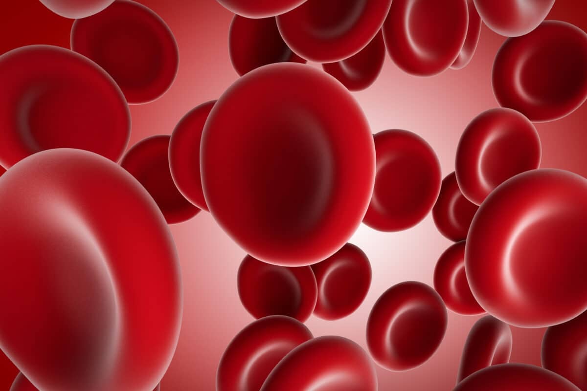
Lymphocytes are a type of white blood cells that play a critical role in your immune system. They are your body’s frontline defenders, targeting harmful invaders like viruses, bacteria, and other pathogens. Produced in the bone marrow, lymphocytes circulate in the bloodstream and reside in lymphatic tissues such as the spleen, lymph nodes, and thymus. Their role is essential in keeping your immune system strong and protecting your body from diseases.

There are three main types of lymphocytes: B-cells, T-cells, and Natural Killer (NK) cells. Each type has a specific function:

  • B-cells produce antibodies, proteins that bind to foreign substances, marking them for destruction.
  • T-cells directly attack infected or cancerous cells and help regulate immune responses.
  • Natural Killer (NK) cells play a role in the early defense against viruses and tumors, recognizing and eliminating compromised cells.

How Do You Measure Lymphocyte Count?

Measuring lymphocyte count involves a straightforward blood test, which provides valuable insights into how well your body is defending against infections and other diseases. Here’s a detailed look at how lymphocyte counts are measured:

Blood Sample Collection

The first step in measuring lymphocyte count is obtaining a blood sample. This is usually done through a venipuncture, where a healthcare professional draws blood from a vein in your arm using a needle. The procedure is quick, generally painless, and only requires a small amount of blood.

Complete Blood Count (CBC) with Differential

Once the blood sample is collected, it is sent to a laboratory for analysis. The most common method for measuring lymphocytes is through a Complete Blood Count (CBC) with a differential. This comprehensive test evaluates the different components of your blood, including:

  • Red Blood Cells (RBCs)
  • White Blood Cells (WBCs)
  • Platelets

The differential analysis portion of the CBC specifically breaks down the white blood cells into their various types, including lymphocytes. Automated machines typically perform this analysis, providing an accurate count of each type of cell present in your blood.

Flow Cytometry

For more detailed and specific information, especially in cases where abnormalities are suspected, flow cytometry in combination with specific antigen labels may be used. This advanced laboratory technique allows for the precise identification and quantification of different lymphocyte subsets, such as:

  • B-Cells
  • T-Cells
  • Natural Killer (NK) Cells

Flow cytometry may help in diagnosing specific conditions, such as certain types of leukemia, lymphoma, and immune system disorders, by providing a more granular view of your lymphocyte populations.

Manual Counting

In some instances, particularly when automated results are inconclusive or when a more detailed examination is necessary, a manual count may be performed. A laboratory technician will examine a blood smear under a microscope to count the lymphocytes manually. While more time-consuming, this method may provide additional insights into the appearance and morphology of the lymphocytes, which may be important for diagnosing specific diseases.

What is a Normal Lymphocyte Count?

A normal lymphocyte count varies depending on age, overall health, and individual factors, but there are general ranges that are considered healthy for most people.

For adults, a normal lymphocyte count typically falls within the following ranges:

  • Absolute Lymphocyte Count: 1,000 to 4,800 lymphocytes per microliter (µL) of blood.

  • Relative Lymphocyte Count: 20% to 40% of the total white blood cell count.

For children, normal lymphocyte counts may be higher, with ranges varying based on age:

  • Infants (up to 12 months): 2,000 to 10,500 lymphocytes per microliter of blood.

  • Toddlers (1-4 years): 2,000 to 9,500 lymphocytes per microliter of blood.

  • Children (5-18 years): 1,250 to 7,000 lymphocytes per microliter of blood.

These ranges serve as general guidelines, and what is considered “normal” may differ slightly depending on the laboratory performing the test. If your lymphocyte count falls outside these ranges, it doesn’t necessarily mean something is wrong, but it may need further testing.

What Does it Mean if You Have High/Low Lymphocyte Count?

Your lymphocyte count may provide important clues about your immune system’s health. Both high and low lymphocyte counts may signal underlying health issues.

High Lymphocyte Count (Lymphocytosis)

A high lymphocyte count, known as lymphocytosis, occurs when there are more lymphocytes in your blood than the normal range. Lymphocytosis may be a temporary response to an infection or a more persistent condition associated with chronic illnesses. Common causes of high lymphocyte counts include:

  • Infections: Viral infections like mononucleosis, hepatitis, and cytomegalovirus (CMV) infection are frequent causes of lymphocytosis. Some bacterial infections, such as in tuberculosis and whooping cough, may also lead to elevated lymphocyte levels.
  • Chronic Inflammatory Conditions: Diseases like rheumatoid arthritis and inflammatory bowel disease (IBD) may cause persistent lymphocytosis due to ongoing inflammation.
  • Lymphocytic Leukemia: A type of blood cancer that begins in the bone marrow, leading to an overproduction of lymphocytes.
  • Stress or Physical Trauma: Acute stress, intense physical activity, or injury may sometimes cause temporary increases in lymphocyte count.

Low Lymphocyte Count (Lymphocytopenia)

A low lymphocyte count, known as lymphocytopenia, occurs when there are fewer lymphocytes in your blood than normal. This may weaken your immune system, making you more susceptible to infections and other health problems. Common causes of low lymphocyte counts include:

  • Viral Infections: Severe viral infections like with HIV may lead to a depletion of lymphocytes over time.
  • Autoimmune Disorders: Conditions such as lupus and multiple sclerosis may result in lymphocytopenia due to the immune system attacking its own cells, including lymphocytes.
  • Bone Marrow Disorders: Diseases that affect bone marrow function, like aplastic anemia or certain cancers, may lead to reduced lymphocyte production.
  • Medications: Certain treatments, like chemotherapy, immunosuppressants, and corticosteroids, may decrease lymphocyte levels as a side effect.
  • Nutritional Deficiencies: Lack of essential nutrients, such as proteins or vitamins, may impair lymphocyte production.

Can Genetics Influence Lymphocyte Count?

Yes, genetics may significantly influence your lymphocyte count. Research has shown that genetic variations may affect how many lymphocytes you have, how they function, and how your immune system responds to various challenges. 

A 2010 study looked at the genetic data of 2,538 people and examined how 2.3 million genetic variations influenced five different types of lymphocytes, including CD4+ T-cells, CD8+ T-cells, and Natural Killer (NK) cells. They found two key genetic regions associated with these lymphocyte levels:

  1. Major Histocompatibility Complex (MHC) Region: This region strongly influences the CD4:CD8ratio, which is important in immune function. The study found two specific genetic variants in the MHC region:
    • One affects levels of CD8+ T-cells (in the class I part of MHC).
    • The other affects levels  of CD4+ T-cells (in the class II part of MHC).
  2. Schlafen (SLFNL) Gene Family: This genetic region is linked to the levels of NK-cells.

The findings suggest that the genetic  variation in the MHC region genes could affect the balance of important immune cells called CD4+ and CD8+ T-cells. These cells help the body to fight off infections. When the balance of these cells is disrupted, it can lead to health problems. For example, some genetic variations in the MHC region are linked to better control of HIV, meaning they might help the immune system handle the virus more effectively. On the other hand, other genetic variations in the MHC region are associated with a higher risk of type 1 diabetes, a condition where the immune system mistakenly attacks the pancreas. These findings show how our genetic variants  can influence our immune system and our risk for certain diseases.

Non-Genetic Factors Influencing Lymphocyte Count

While genetic plays a significant role in determining your lymphocyte count, several non-genetic factors may also influence these crucial immune cells. These factors may cause fluctuations in lymphocyte levels and impact your overall immune health.

1. Infections

Infections are one of the most common non-genetic factors affecting lymphocyte count. Viral infections, in particular, may cause significant changes:

  • Viral Infections: Conditions like the flu, mononucleosis, and HIV may lead to lymphocytosis (increased lymphocyte count) as your body ramps up its immune response to fight the virus.
  • Bacterial Infections: Some bacterial infections, such as tuberculosis, may also affect lymphocyte levels, though typically less dramatically than viral infections.

On the flip side, chronic viral infections, such as with HIV, may lead to lymphocytopenia (decreased lymphocyte count) over time, weakening the immune system.

2. Autoimmune Disorders

Autoimmune diseases occur when the immune system mistakenly attacks the body’s own tissues. This may have a profound impact on lymphocyte count:

  • Lupus: In lupus, an autoimmune condition, lymphocyte counts may be abnormally low due to the immune system’s dysregulation and the potential impact of treatments like corticosteroids.
  • Rheumatoid Arthritis: This chronic inflammatory disorder often causes elevated lymphocyte counts as the immune system remains in a state of persistent activation.

3. Medications and Treatments

Certain medications and medical treatments may significantly influence lymphocyte count:

  • Chemotherapy: Used to treat cancer, chemotherapy may reduce lymphocyte counts, making patients more susceptible to infections.
  • Immunosuppressants: Drugs used to prevent organ rejection in transplant patients, or to treat autoimmune diseases, may lower lymphocyte counts, dampening the immune response.
  • Corticosteroids: These anti-inflammatory drugs, synthetic versions of cortisol,  may reduce lymphocyte numbers by altering the distribution and production of these cells in the body.

4. Stress

Both physical and psychological stress may impact lymphocyte count:

  • Acute Stress: Short-term physical stress, such as intense exercise or injury, may temporarily increase lymphocyte levels as part of the body’s immediate response to perceived threats.
  • Chronic Stress: Prolonged psychological stress may lead to a decrease in lymphocyte count, weakening the immune system and increasing susceptibility to infections.

5. Nutritional Status

Your diet and nutritional status play a crucial role in maintaining healthy lymphocyte levels:

6. Age

Lymphocyte count naturally changes with age:

  • Children: Typically have higher lymphocyte counts than adults due to their developing immune systems.
  • Elderly: As people age, lymphocyte production may decrease, leading to lower counts and a weakened immune response, making older adults more susceptible to infections.

7. Lifestyle Factors

Certain lifestyle choices may also influence lymphocyte count:

  • Smoking: Smoking has been linked to both elevated and decreased lymphocyte counts, depending on the stage of exposure and overall health of the smoker.
  • Alcohol Consumption: Excessive alcohol intake may impair immune function and reduce lymphocyte count, increasing the risk of infections.
  • Exercise: Regular, moderate exercise generally supports a healthy immune system and stable lymphocyte counts, while extreme exercise, like marathon running, may temporarily lower lymphocyte levels.

8. Environmental Exposure

Exposure to certain environmental factors may impact lymphocyte count:

  • Pollutants: Long-term exposure to environmental pollutants, such as heavy metals and industrial chemicals, may affect lymphocyte levels and compromise immune function.
  • Radiation: Exposure to high levels of radiation, whether from medical treatments or environmental sources, may reduce lymphocyte counts and damage the immune system.

While your genetic makeup sets the foundation for your lymphocyte count, non-genetic factors may significantly shape its daily fluctuations. By understanding and addressing these influences—such as stress, diet, and environmental exposures—you may actively support your immune system. Taking proactive steps to maintain a healthy lifestyle and regularly monitoring your lymphocyte count, particularly if you’re managing infections, autoimmune conditions, or undergoing treatments like chemotherapy, may empower you to optimize your immune health.

Summary:

  • Lymphocytes are crucial white blood cells that help protect the body from infections and diseases.
  • They are measured through blood tests such as the Complete Blood Count (CBC) with a differential.
  • Normal lymphocyte counts range from 1,000 to 4,800 per microliter in adults, with higher levels in children.
  • Elevated lymphocyte counts may indicate infections, chronic inflammatory diseases, or hematological cancers.
  • Reduced lymphocyte counts may result from viral infections, autoimmune diseases, bone marrow disorders, or adverse effects from certain medications.
  • Genetic factors play a significant role in determining lymphocyte levels and function.
  • Non-genetic factors affecting lymphocyte counts include infections, autoimmune conditions, treatments like chemotherapy and immunosuppression, stress (physical and psychological), nutritional deficiencies, age, lifestyle choices (smoking, alcohol consumption), and environmental exposures (pollutants, radiation).

References:

  1. https://pubmed.ncbi.nlm.nih.gov/11196672/

  2. https://www.ncbi.nlm.nih.gov/pmc/articles/PMC8546033/

  3. https://www.nature.com/articles/6363702 

  4. https://www.medicalnewstoday.com/articles/320987

  5. https://www.sciencedirect.com/topics/biochemistry-genetics-and-molecular-biology/lymphocyte-count

  6. https://www.nature.com/articles/jhg20122

  7. https://www.sciencedirect.com/topics/agricultural-and-biological-sciences/lymphocyte

  8. https://www.britannica.com/science/human-genetics

  9. https://my.clevelandclinic.org/health/body/24630-t-cells

  10. https://www.healthcare.uiowa.edu/path_handbook/appendix/heme/pediatric_normals.html

  11. https://pubmed.ncbi.nlm.nih.gov/20045101/

Can Genes Influence Your Metamyelocyte Count

Man lying on the couch looking tired, reflecting the potential impact of genetic factors on metamyelocyte count

Disclaimer: This article is for informational purposes only and is not intended to diagnose any conditions. LifeDNA does not provide diagnostic services for any conditions mentioned in this or any other article.

Have you ever received a blood test result that included a term you didn’t recognize, like “metamyelocyte count”? While it may sound intimidating, understanding this may provide valuable insights into your overall health. Recognizing its significance may help you gain insight into your body’s response to stress and disease, which may potentially lead to more informed decisions about your health care.

What is Metamyelocyte Count?

Metamyelocytes are immature neutrophils in the early stages of white blood cell development, which play an important role in your immune system by helping your body fight infections. In a healthy individual, metamyelocytes are typically confined to the bone marrow where blood cells are produced and do not appear in the bloodstream. However, under certain conditions—such as infection, inflammation, or stress—the body may release immature white blood cells, including metamyelocytes, into the bloodstream.

The metamyelocyte count refers to the number of these immature cells found in a blood sample. In a routine blood test, metamyelocytes are not usually present in the blood. If they are detected, it might suggest that the bone marrow is producing white blood cells rapidly in response to an issue, such as an infection or another underlying condition. Doctors use the metamyelocyte count as part of a broader blood test to help diagnose and monitor various health conditions.

How Do You Measure Metamyelocyte Count?


The metamyelocyte count is measured as part of a routine blood test called a complete blood count (CBC) with a differential (including a breakdown of different white blood cell types). This test provides doctors with a detailed overview of your blood’s composition, particularly focusing on the various types of cells present, including those involved in your immune response.


Here’s what you can expect if your doctor orders this test:


  • Getting a Blood Sample: A small amount of blood is drawn, usually from a vein in your arm. This process is quick and typically done during a visit to your doctor or at a lab.
  • Analyzing the Blood: Once your blood sample reaches the lab, it iss analyzed to determine the types and quantities of cells present. The lab specifically looks for any immature white blood cells, such as metamyelocytes, which are not normally found in circulating blood.
  • Understanding the Results: Normally, metamyelocytes are only found in the bone marrow, not in the bloodstream. If they show up in your blood, it could be a sign that your body is responding to a particular condition, such as an infection, inflammation, or another type of stress.
  • What the Results Mean for You: If your metamyelocyte count is higher than normal, your doctor might look into other tests or ask about symptoms you’re experiencing. This helps them figure out if there’s an underlying condition that needs attention.

What is the Normal Metamyelocyte Count?

While genetic factors play a role in the normal proportions of various blood cells, the specific genetic variants  influencing metamyelocyte counts are not well-defined. However,  the normal levels of metamyelocytes may vary. While their presence in the bloodstream is generally abnormal, their presence in the bone marrow is part of the normal blood cell maturation. Understanding this helps in assessing whether their presence indicates a health issue.

Presence in Blood

The normal metamyelocyte count is zero when measured in the bloodstream. If metamyelocytes are detected in the bloodstream, it is generally considered abnormal and may indicate that the body is responding to an underlying condition. The presence of metamyelocytes in the blood may need further examination to determine the cause and assess the need for treatment.

In Bone Marrow

In the bone marrow, metamyelocytes are a normal part of the development process for white blood cells. While there isn’t a specific “normal” count for metamyelocytes in the bone marrow due to variations in individual health and age, their presence is expected as they mature into more specialized white blood cells. The bone marrow typically contains a range of immature white blood cells, including metamyelocytes, which are part of the normal hematopoiesis (blood cell formation) process.

What Causes High or Low Metamyelocyte Count?

High Metamyelocyte Count

A high metamyelocyte count means that there are more of these immature white blood cells in your bloodstream than usual. This is not typical, as metamyelocytes should normally only be found in the bone marrow. Elevated levels can suggest several potential issues:

  • Infection: The body may release immature white blood cells into the bloodstream while fighting an infection.
  • Inflammation: Chronic inflammation or conditions causing ongoing stress to the body may lead to increased metamyelocyte production.
  • Bone Marrow Disorders: Conditions like chronic myelogenous leukemia (CML), myelodysplastic syndromes (MDS), or other myeloproliferative disorders may lead to an increased release of immature cells, including metamyelocytes, into the bloodstream.
  • Severe Physical Stress: Physical stress, severe illness, or trauma can also cause a temporary increase in metamyelocyte count as the body mobilizes its immune defenses.
  • Toxic Exposure: Exposure to certain toxins, chemotherapy, or radiation may damage the bone marrow, leading to an increased release of immature cells.

Low Metamyelocyte Count

A low metamyelocyte count is rarely discussed because metamyelocytes are typically not present in the bloodstream under normal conditions. Their presence is expected primarily in the bone marrow, where they develop as part of the neutrophil maturation process. However, if their count is unusually low in the bone marrow, it may signal an underlying issue affecting bone marrow function.

For example, aplasia or dysfunction of the bone marrow can impair the production of metamyelocytes and other essential blood cells.

  • Aplastic Anemia: Aplastic anemia is a serious condition in which the bone marrow fails to produce enough blood cells, including white blood cells (WBCs) and their precursors, such as metamyelocytes. In this scenario, a low metamyelocyte count reflects the marrow’s inability to generate and release sufficient immune cells. This deficit weakens the body’s immune response, leaving it vulnerable to infections and other complications.
  • Certain Types of Leukemia: In some forms of leukemia, particularly those where the bone marrow becomes crowded with abnormal, cancerous cells, the production of normal blood cells is significantly hindered. As a result, the number of healthy precursor cells, including metamyelocytes, may be lower than expected. A bone marrow biopsy in these cases often reveals a diminished count of metamyelocytes, indicating that the marrow’s capacity to produce normal blood cells is compromised.
  • Chronic Diseases: Long-standing chronic illnesses, such as autoimmune diseases, chronic infections, or severe inflammatory disorders, can lead to bone marrow suppression. This results in a reduced production of all blood cell types, including neutrophil precursors like metamyelocytes.
  • Malnutrition: Nutritional deficiencies, particularly those affecting vitamins and minerals essential for blood cell production (e.g., vitamin B12, folate, or iron), can impair the bone marrow’s ability to produce white blood cells. In cases of severe malnutrition, a bone marrow examination may reveal fewer immature cells, including metamyelocytes.

When Should Metamyelocytes Be Detected in Blood?

Metamyelocytes should not typically be present in peripheral blood. However, they may appear in the bloodstream under certain conditions, such as:

  • Severe Infections: When the body requires an urgent response to infection, the bone marrow may release immature cells like metamyelocytes into the blood.
  • Hematologic Disorders: Conditions affecting the bone marrow, such as leukemia or myeloproliferative disorders, may result in the release of metamyelocytes into circulation.

Immature Neutrophils as Indicators of Sepsis 

Myelocytes and metamyelocytes, as well as other immature neutrophils are emerging as important biomarkers for assessing infection severity and predicting outcomes. Band cells, another type of immature neutrophil, signal severe infections like sepsis when present in high numbers—a condition known as a “shift to the left.” Elevated band cell levels are linked to poorer sepsis outcomes. 

Research has demonstrated that high percentages of band cells are associated with poorer outcomes in sepsis. Specifically, a groundbreaking study found that band cells had a sensitivity of 84% and a specificity of 71% for detecting definite sepsis at an 8.5% cutoff point. This makes band cells a valuable tool for differentiating between sepsis and non-infectious systemic inflammatory response syndrome (N-I SIRS).

Myelocytes and metamyelocytes, though less commonly monitored, also provide critical prognostic information. The same study discovered that patients with up to 40% of these cells in their blood had significantly higher mortality rates. Specifically, patients who died within the first week of ICU stay had a median of 9% myelocytes and metamyelocytes in their blood, compared to much lower levels in those who survived longer.

The presence of immature neutrophils in the blood, particularly in ICU settings, is associated with higher mortality risk, highlighting their potential as valuable markers in managing severe infections. Monitoring these cells aids in distinguishing between systemic inflammatory response syndrome (SIRS) and sepsis, thus guiding treatment and improving patient management.

Non-Genetic Factors Influencing Metamyelocyte Count

Several non-genetic factors can influence the metamyelocyte count in your blood. These factors often relate to the body’s response to various conditions or stressors. Here’s how they might affect your metamyelocyte levels:

  • Infections: During infections, especially the severe ones with bacterial origin like sepsis, the body releases immature white blood cells, such as metamyelocytes, into the bloodstream. This response is part of the immune system’s effort to rapidly combat the invading pathogens.
  • Inflammation: Chronic inflammation or inflammatory diseases such as rheumatoid arthritis or inflammatory bowel disease, can stimulate the bone marrow to produce more white blood cells, including metamyelocytes, as the body attempts to manage and repair affected tissues.
  • Stress: Physical or emotional stress can lead to an increase in the production of white blood cells, including metamyelocytes, as part of the body’s heightened immune response. For example, severe trauma or surgery can temporarily elevate metamyelocyte counts.
  • Bone Marrow Disorders: Disorders affecting the bone marrow, such as leukemia or myelodysplastic syndromes, can result in the abnormal production and release of immature cells like metamyelocytes into the bloodstream. In acute myeloid leukemia (AML), this process is particularly pronounced, leading to elevated metamyelocyte levels.
  • Medications: Certain medications, such as granulocyte colony-stimulating factor (G-CSF) used during chemotherapy, can stimulate bone marrow activity and increase metamyelocyte counts as they enhance white blood cell production.
  • Nutritional Deficiencies: While nutritional deficiencies are less directly linked to metamyelocyte counts, they can impact overall blood cell health. Deficiencies in essential nutrients like vitamin B12 or folate may indirectly affect white blood cell production by impairing bone marrow function.
  • Systemic Diseases: Systemic diseases, such as chronic kidney disease or liver disease, can alter bone marrow function and lead to changes in blood cell counts, including metamyelocytes. Chronic kidney disease, for instance, may influence the production of immature cells in the blood.


Summary


  • Metamyelocytes are immature white blood cells in early stages of development, maturing into neutrophils which play a crucial role in the immune system by fighting infections.
  • These immature cells are normally found only in the bone marrow.
  • The metamyelocyte count is assessed via a complete blood count (CBC) with a differential, which provides a detailed breakdown of different white blood cell types. Typically, metamyelocytes are absent from the bloodstream.
  • In a healthy individual, the normal metamyelocyte count in the blood is zero. Their presence in the bloodstream suggests a potential health issue and may necessitate further investigation.
  • In the bone marrow, metamyelocytes are a normal part of the blood cell maturation process, with no specific “normal” count due to variability among individuals.
  • A high metamyelocyte count can indicate various issues, including infections, chronic inflammation, bone marrow disorders, severe physical stress, or toxic exposure. Elevated levels, especially in blood,  are important for diagnosing and monitoring health conditions.
  • A low metamyelocyte count is less frequently discussed but may suggest bone marrow problems or chronic diseases affecting white blood cell production.
  • Metamyelocytes, along with other immature neutrophils like band cells, are used to assess the severity of infections and patient prognosis. High levels of band cells often signal severe infections such as sepsis and can differentiate between sepsis and non-infectious systemic inflammatory response syndrome (N-I SIRS).
  • Elevated myelocyte and metamyelocyte counts are associated with poorer outcomes in ICU settings and can help predict patient prognosis. High levels correlate with a higher risk of mortality.

References

  1. https://imagebank.hematology.org/image/60398/metamyelocyte 
  2. https://www.sciencedirect.com/topics/immunology-and-microbiology/metamyelocyte 
  3. https://www.sciencedirect.com/topics/agricultural-and-biological-sciences/metamyelocyte 
  4. https://www.biron.com/en/glossary/metamyelocyte/ 
  5. https://www.ncbi.nlm.nih.gov/pmc/articles/PMC4355545/
  6. https://www.ncbi.nlm.nih.gov/pmc/articles/PMC8081893/#B79
  7. https://karger.com/mpp/article-abstract/16/5/344/203779/The-Diagnostic-Value-of-Absolute-Neutrophil-Count 


Albumin Level And What It Means For Your Health

Close-up image of nerve cells highlighting the connection between albumin levels and vascular health

Disclaimer: This article is for informational purposes only and is not intended to diagnose any conditions. LifeDNA does not provide diagnostic services for any conditions mentioned in this or any other article.


Albumin, a protein produced by the liver, plays an important role in maintaining our overall health. This protein is the most abundant circulating protein in plasma, representing about half of the total protein content (3.5 g/dL to 5 g/dL) in healthy individuals. Albumin’s primary functions include modulating plasma oncotic pressure and transporting various substances within the blood. 

By understanding albumin’s functions, we can better appreciate how our body works to stay healthy and how certain health conditions might be linked to albumin levels.

What is Albumin?

Albumin is a type of protein produced by the liver and released into the bloodstream. It consists of 585 amino acids, the building blocks of proteins, connected together in a small, globular structure. The liver produces about 10 to 15 grams of albumin daily, which is then rapidly excreted into the bloodstream. While only a small amount of albumin is stored in the liver, it is mostly found in the bloodstream, as well as in the fluid between cells, and in other body fluids. In clinical practice, doctors measure serum albumin levels through standard blood or urine tests to assess a person’s nutritional status and overall health.

What is The Function of Albumin in Our Body? 

Albumin is a multifunctional protein essential for various physiological processes. Its diverse functions range from regulating fluid balance to transporting vital substances and contributing to several biochemical activities within the body. The following sections detail these important functions.

Modulating Plasma Oncotic Pressure

Albumin is the most significant modulator of plasma oncotic pressure, which helps maintain the balance of fluid between blood vessels and tissues. By influencing oncotic pressure, albumin prevents fluid leakage into the extravascular space, thus maintaining blood volume and pressure.

Transporting Substances

Albumin acts as a carrier for various endogenous and exogenous substances. These substances, also known as ligands, include bilirubin, ions, fatty acids, hormones, and drugs such as methadone, propranolol, thiopental, furosemide, warfarin, and methotrexate. Albumin’s binding capacity helps reduce the toxicity of these substances and facilitates their transport to target sites within the body.

Other Functions

Apart from modulating oncotic pressure and transporting substances, albumin also plays a role in maintaining acid-base balance, acting as an antioxidant, and serving as a buffer in blood plasma. Furthermore, albumin is involved in binding and transporting hormones such as thyroxine, cortisol, and testosterone, as well as contributing to calcium homeostasis by binding a significant portion of circulating calcium.

Genetic Factors Influencing Albumin Levels

A study looked at whether low levels of albumin (a protein in the blood) may increase the risk of developing high blood pressure (hypertension). The researchers used data from a large group of people in Korea and found that people with low albumin were more likely to develop high blood pressure over time. They also discovered two genetic markers that seem to be linked to both low albumin levels and higher blood pressure. These genetic markers could affect how blood vessels work, which might explain why low albumin can lead to high blood pressure. While the study has some limitations, like small sample sizes and potential biases, it suggests that low albumin could be an important factor in predicting hypertension, and more research is needed to fully understand how it works.

This study shows that low albumin levels might not just be a sign of poor nutrition or liver issues, but may also be an early warning sign for developing high blood pressure (hypertension). It showed that low albumin may affect how your blood vessels function, which might contribute to the development of hypertension, a major risk factor for heart disease and stroke. These genetic studies reveal that albumin levels are not just influenced by dietary and environmental factors but also by genetic variations. Understanding these genetic influences may help improve diagnosis and treatment by identifying specific genetic loci associated with abnormal albumin levels, leading to better diagnostic tools and personalized treatments for conditions like liver disease and chronic kidney disease. 


What is the Normal Range of Albumin?

The normal range of serum albumin in the blood is between 3.4 to 5.4 g/dL (34 to 54 g/L). However, normal value ranges may slightly vary among different laboratories. Maintaining albumin levels within this range is crucial for the body’s homeostasis and optimal functioning.

What is Hypoalbuminemia?

Hypoalbuminemia refers to lower-than-normal levels of serum albumin. This condition can be a sign of various underlying health issues, including:

Liver Diseases: Hypoalbuminemia is often linked to liver diseases such as hepatitis and cirrhosis. The liver is the primary site of albumin production, so any condition impairing liver function can result in decreased albumin synthesis. In hepatitis, inflammation of the liver cells can hinder albumin production. Cirrhosis, which is characterized by scarring of liver tissue, disrupts normal liver function and significantly reduces albumin production. Consequently, hypoalbuminemia may be an indicator of compromised liver health.

Kidney Diseases: A kidney disease particularly nephrotic syndrome may lead to hypoalbuminemia due to excessive loss of albumin in the urine. Nephrotic syndrome damages the kidney’s filtering units, allowing large amounts of albumin to leak into the urine. This significant loss of albumin from the bloodstream lowers the overall albumin levels, leading to hypoalbuminemia. Chronic kidney disease and other glomerular disorders may possibly cause albuminuria.

<You may also like: Can Genetics Increase Your Risk for Kidney Stones

Malnutrition and Nutrient Absorption Issues: Malnutrition and conditions affecting nutrient absorption, such as celiac disease, Crohn’s disease, and Whipple disease, can result in hypoalbuminemia. These conditions impair the digestive system’s ability to absorb proteins and other nutrients essential for albumin production. Prolonged malnutrition leads to insufficient protein intake, directly impacting the liver’s ability to produce adequate amounts of albumin, thus causing hypoalbuminemia.

Increased Albumin Loss: Hypoalbuminemia can also occur due to increased albumin loss in various situations. Severe burns can cause extensive protein loss through damaged  skin. Protein-losing enteropathies, which involve the loss of proteins from the gastrointestinal tract, can significantly reduce serum albumin levels. Additionally, critical illnesses that increase vascular permeability allow albumin to escape from blood vessels into tissues, further depleting serum albumin levels and contributing to hypoalbuminemia.

How to Treat Hypoalbuminemia

Hyperalbuminemia is typically managed by addressing the underlying cause, such as rehydration in cases of dehydration. Monitoring and adjusting dietary protein intake may also be necessary in some cases. The management of hypoalbuminemia involves addressing the underlying cause. Treatment strategies may include:

Nutritional Support: Ensuring adequate protein intake is vital. This involves incorporating more protein-rich foods such as meat, fish, eggs, and legumes into the diet or using supplements. Addressing malnutrition and conditions that affect nutrient absorption, like celiac disease or Crohn’s disease, is crucial. A dietitian can help create a suitable meal plan to meet the patient’s nutritional needs.

Medications: Managing the underlying conditions, such as liver or kidney  diseases, is essential. For liver diseases, antiviral drugs or anti-inflammatory medications might be necessary. For kidney diseases like nephrotic syndrome, corticosteroids or other immunosuppressive drugs can help reduce protein loss in urine.


Fluid Management: In severe hypoalbuminemia cases, administering albumin or other colloid fluids can help maintain plasma oncotic pressure and prevent edema. This treatment is typically provided in a hospital setting under careful monitoring. Diuretics may also be used to manage fluid retention, but they must be used cautiously to avoid worsening the condition.

What is Hyperalbuminemia?

Hyperalbuminemia, characterized by higher-than-normal levels of serum albumin, is less common but can occur due to several factors. Primarily, dehydration is the most common cause, as fluid loss concentrates albumin levels. Additionally, consuming a high protein diet can elevate albumin levels by stimulating the liver to produce more. Another contributing factor is the prolonged application of a tourniquet during blood sampling, which can artificially increase albumin blood levels. Similarly, conditions like insulin resistance within metabolic syndrome can trigger the liver to increase albumin production.

How Do You Treat Hyperalbuminemia?

Hyperalbuminemia is typically managed by addressing the underlying cause. Treatment strategies may include:

Rehydration: Dehydration is a common cause of hyperalbuminemia. Rehydration through oral fluids or intravenous solutions can help restore fluid balance and normalize albumin levels​​​​. Severe dehydration is usually treated with fluids and zinc supplements. Zinc helps decrease cell swelling caused by low water intake and promotes salt retention​

Dietary Adjustments: Reducing the intake of protein-rich foods can help manage elevated albumin levels, especially when these levels are linked to an excessive protein diet.

Managing Underlying Conditions: Addressing underlying conditions like metabolic syndrome is crucial. This involves adopting a healthier diet, increasing physical activity, and using medications to control insulin resistance and related health issues.

Correcting Sampling Techniques: Avoiding prolonged application of a tourniquet during blood sampling can prevent artificially elevated albumin blood levels.

Diagnosing and Testing Albumin Levels 

Diagnosing and testing serum albumin levels can be accomplished through several methods, each serving to evaluate the concentration of albumin in the blood or other body fluids and identify potential underlying health issues. These methods include:

Serum Albumin Test

The most common test, which measures the amount of albumin in the blood. A healthcare provider takes a blood sample, typically from a vein in the arm, and the sample is analyzed in a laboratory. This test helps in assessing liver function, kidney function, and nutritional status.

Urine Albumin Test

Also known as the microalbumin test, this measures the level of albumin in the urine to identify early kidney damage, particularly in people with diabetes or high blood pressure. This test can be conducted using a single urine sample or a 24-hour urine collection.

Albumin/Creatinine Ratio (ACR)

The ACR test assesses the amount of albumin relative to creatinine in a urine sample. This ratio provides a more precise evaluation of kidney function and helps detect kidney disease at an early stage.

Liver Function Tests

A series of blood tests that measure albumin levels along with various liver enzymes and proteins to assess overall liver health. 

Comprehensive Metabolic Panel (CMP)

A broader test that includes measuring albumin levels along with other electrolytes, proteins, and liver enzymes. It provides a comprehensive overview of metabolic and organ functions, including liver and kidney health.

Some FAQs on Albumin

What If Your Albumin Is High During Pregnancy

During pregnancy, especially in the second and third trimesters, it is common to see a decrease in serum albumin levels due to increased blood volume and fluid retention. High albumin levels during pregnancy could indicate dehydration or other underlying health issues and should be discussed with a healthcare provider.

Does Alcohol Affect Albumin?

Chronic alcohol consumption may lead to liver damage, impairing the liver’s ability to produce albumin. This can result in lower serum albumin levels and contribute to conditions like hypoalbuminemia.

Does Diet and Lifestyle Affect Albumin?

Diet and lifestyle significantly impact albumin levels. Adequate protein intake is essential for maintaining normal albumin levels. Malnutrition, poor dietary habits, and conditions that affect nutrient absorption may lead to decreased albumin production.

Does Albumin Increase with Age?

Albumin levels tend to decrease with age due to various factors such as reduced liver function, changes in body composition, and increased prevalence of chronic diseases. Monitoring albumin levels in older adults is important for assessing their nutritional status and overall health.

Tips to Naturally Have Balanced/ Normal Range of Albumin Levels

Maintaining balanced and normal albumin levels is crucial for overall health. Here are some tips to naturally achieve this:

Ensure Adequate Protein Intake: Consuming sufficient amounts of protein is essential.This helps the liver produce adequate amounts of albumin.

Stay Hydrated: Dehydration can lead to elevated albumin levels. Make sure to drink at least 8 glasses of water daily, or more if you are physically active or in a hot climate.

Avoid Excessive Alcohol Consumption: Excessive alcohol intake can damage the liver, affecting its ability to produce albumin. Limit alcohol consumption to moderate levels.

References

  1. https://www.mountsinai.org/health-library/tests/albumin-blood-serum-test#:~:text=Albumin%20is%20a%20protein%20made,be%20measured%20in%20the%20urine.
  2. https://www.ncbi.nlm.nih.gov/books/NBK459198/
  3. https://my.clevelandclinic.org/health/diseases/22529-hypoalbuminemia
  4. https://my.clevelandclinic.org/health/articles/22243-amino-acids
  5. https://www.sciencedirect.com/science/article/abs/pii/S0304416513001207
  6. https://academic.oup.com/clinchem/article/43/10/2014a/5640631
  7. https://www.sciencedirect.com/science/article/abs/pii/B9780080196039500120
  8. https://www.nature.com/articles/s41598-018-33072-z
  9. https://www.frontiersin.org/journals/genetics/articles/10.3389/fgene.2024.1372042/full

White Blood Cell: Immunity, Genetic Basis And More

Disclaimer: This article is for informational purposes only and is not intended for diagnostic use. LifeDNA does not provide diagnostic reports on any traits discussed. Genetics is just one piece of the puzzle; please consult a healthcare professional for comprehensive guidance on any health condition.

White blood cells (WBCs), or leukocytes, are crucial for the immune system. They circulate in the blood and defend the body against infections, foreign invaders, and diseases. There are several types of WBCs, including lymphocytes, monocytes, neutrophils, eosinophils, and basophils, each with specialized functions. 

Neutrophils, for instance, are the most abundant and are first responders to infection sites, while lymphocytes, such as B and T cells, are central to the adaptive immune response. The presence and activity of WBCs are vital for maintaining overall health and preventing infections.

Why are WBCs Important?

WBCs are essential for maintaining the immune defense system. They play a pivotal role in protecting the body from infections by identifying and destroying pathogens such as bacteria, viruses, and fungi. WBCs also help remove dead or damaged cells and respond to allergens and other foreign substances. 

Their ability to move to infection sites and engulf or neutralize harmful invaders is crucial for preventing the spread of disease and aiding in the healing process. Without adequate WBCs, the body becomes susceptible to infections and diseases, highlighting their importance in overall health and immune function.

What Do Low and High WBC Counts Mean?

Low WBC counts, or leukopenia,  implies a weak immune system and increased infection susceptibility. Causes of low WBC counts include bone marrow disorders, autoimmune diseases, severe illnesses, certain medications, and chemotherapy. Conversely, high WBC counts, known as leukocytosis, can suggest the presence of an infection, inflammation, stress, or other conditions such as leukemia. Elevated WBC levels might also result from intense physical activity or an immune response to trauma. Abnormally low and high WBC counts require further medical evaluation to determine the underlying cause and appropriate treatment.

Genetics of WBC Count: A Brief Review

DARC gene

A 2011 genome-wide association study (GWAS) involving 16,388 people from the African American population showed that members of this population typically have lower overall WBC and neutrophil counts due to the “null” variant of the Duffy Antigen Receptor for Chemokines (DARC) gene. Interestingly, this variant also protects against some forms of malaria. The DARC gene is significant for its impact on blood type classification and its role in immune function;,with variations in this gene influencing variations in WBC counts and susceptibility to diseases. The study further confirms that also the CXCL2, CDK6, and PSMD3-CSF3 gene regions are influential contributors to WBC counts across different populations, emphasizing their broader relevance.


A 2012 GWAS involved 13,923 participants from both African ancestry and European ancestry. This study also found a significant association between WBC count and the DARC gene in the participants with African ancestry. It validated the association between WBC levels and the regulatory SNP variant rs2814778, which causes the Duffy negative phenotype (Fy-/-). Additionally, the study identified interaction effects of two variants, rs2814778 and rs12075, which are responsible for the differing antigens in the Duffy blood group system.

Other Studies

A 2019 GWAS involving 2064 discovery and 407 replication patients identified a new genetic locus at 14q24.3 to be associated with WBC counts. The 14q24.3 locus was also associated with acute stroke outcome and the expression of the ACOT1 and PTGR2 genes.


A 2021 meta-analysis of GWAS involving 17,802 participants of African ancestry highlights the significant genetic influence on WBC and their subtype counts. The study identified five novel loci: LOC126987/MTCO3P14, LINC01525, GAPDHP32/HSD3BP3, FLG-AS1/HMGN3P1, and TRK-CTT13-1/MGST3. 

Genetic Connection with Asthma

Asthma is a chronic respiratory condition characterized by inflammation and narrowing of the airways, leading to difficulty in breathing. The genetic connection between WBC count and asthma is an area of significant research interest. Studies have shown that specific genetic variants associated with WBC count also influence the risk of developing asthma. Some genes, such as those located in the 17q21 region (e.g., GSDMA, MED24, PSMD3), have been associated with variations in WBC count and an increased risk of asthma. These genes are involved in immune system regulation and inflammation, important in WBC function and asthma pathogenesis.

The genetic variants that affect WBC count often impact inflammatory pathways. Since asthma is a chronic inflammatory disease of the airways, the overlap in genetic determinants suggests that individuals with specific genetic profiles may be predisposed to a certain inflammatory response type which influences WBC levels and asthma severity.

Non-Genetic Factors Influencing WBC Count

Non-genetic factors significantly influence WBC counts. Infections are a primary factor, typically causing an increase in WBCs as the body fights off pathogens. Chronic diseases, such as autoimmune disorders and cancer, can also alter WBC levels. Medications, including chemotherapy and certain antibiotics, can suppress WBC production. 

Lifestyle factors like stress, smoking, diet, and physical activity also impact WBC count. Acute physical or emotional stress can cause temporary increases, while chronic stress can lead to longer-term changes. Nutritional deficiencies, particularly in vitamins like B12 and folate, can also affect WBC production and function.

Optimizing Your WBC Levels

WBCs play a crucial role in your immune system, defending your body against infections and diseases. Maintaining optimal WBC levels is essential for overall health. Here are some strategies to help you optimize your WBC levels:

1. Balanced Diet

  • Nutrients: Ensure your diet includes plenty of vitamins and minerals, particularly vitamin C, vitamin E, zinc, and folic acid, which support immune function.
  • Food: Incorporate a variety of fruits, vegetables, lean proteins, and whole grains. Food items such as citrus fruits, berries, nuts, seeds, and leafy greens are particularly beneficial.

2. Regular Exercise

  • Moderate Exercise: Engage in regular, moderate exercise such as walking, jogging, or swimming. Exercise can boost your immune system by promoting healthy circulation, which allows WBCs to move more efficiently throughout your body.

3. Adequate Sleep

  • Sleep Quality: Aim for 7-9 hours of quality sleep per night. Sleep is crucial for the production and function of WBCs. Poor sleep can weaken your immune response and reduce your WBC count.

4. Stress Management

  • Techniques: Practice stress-reducing techniques such as meditation, yoga, deep breathing exercises, or hobbies you enjoy. Chronic stress can negatively impact your immune system and WBC levels.

5. Hydration

  • Water Intake: Stay adequately hydrated by drinking plenty of water throughout the day. Proper hydration supports overall bodily functions, including the production of WBCs.

6. Avoiding Infections

  • Hygiene: Maintain good hygiene practices, such as regular handwashing and avoiding contact with sick individuals, to reduce your risk of infections that can deplete WBCs.

7. Medical Guidance

  • Regular Check-Ups: Schedule regular health check-ups to monitor your WBC levels and overall health. Consult with your healthcare provider if you have concerns about your WBC count or immune function.

8. Avoiding Harmful Substances

  • Substance Use: Avoid smoking and limit alcohol consumption. Both smoking and excessive alcohol intake can weaken your immune system and negatively affect WBC production.

By incorporating these strategies into your lifestyle, you can help maintain optimal WBC levels and support your immune system. Always consult with a healthcare professional before making significant changes to your diet or exercise routine, especially if you have existing health conditions.

References

  1. https://www.cancer.gov/publications/dictionaries/cancer-terms/def/white-blood-cell#
  2. https://journals.plos.org/plosgenetics/article?id=10.1371/journal.pgen.1002108 
  3. https://pubmed.ncbi.nlm.nih.gov/22037903/
  4. https://www.ahajournals.org/doi/10.1161/STROKEAHA.119.026593
  5. https://www.frontiersin.org/journals/genetics/articles/10.3389/fgene.2021.749415/full

Aspartate Aminotransferase: The Blood Marker & Genetic Influence

Black and white image of a muscular man, symbolizing the connection between muscle health and aspartate aminotransferase (AST) levels

Disclaimer: This article is for informational purposes only and is not intended for diagnostic use. LifeDNA does not provide diagnostic reports on any traits discussed. Genetics is just one piece of the puzzle; please consult a healthcare professional for comprehensive guidance on any health condition.

Aspartate aminotransferase (AST) is also known as serum glutamic-oxaloacetic transaminase (SGOT). It is an enzyme found mainly in the liver and heart. It is also in smaller amounts in tissues such as muscles, kidneys, and the brain. 

AST plays a vital role in the amino acid metabolism process. It speeds up a reaction where an amino group (-NH2) is moved from one molecule (aspartate) to another (alpha-ketoglutarate), creating two new molecules (oxaloacetate and glutamate). This reaction is part of a crucial metabolic reaction in the body called the citric acid cycle. This cycle is the main way by which cells produce energy.

AST is also one of the four liver enzymes. The others are alkaline phosphatase (ALP), alanine transaminase (ALT), and gamma-glutamyl transferase (GGT). Liver enzymes are proteins that facilitate specific chemical reactions in the liver.

AST concentration in the blood is commonly measured in liver function tests to diagnose and monitor liver health and to identify conditions such as hepatitis, cirrhosis, and liver fibrosis. AST levels also assess muscle damage, heart attacks, and other medical conditions that can lead to cellular injury.

The Four Liver Enzymes

The four liver enzymes include ALT, AST,  ALP, and GGT. ALT and AST are involved in amino acid metabolism. They are released into the bloodstream whenever the liver cells are damaged. ALP is associated with bile duct function. It is elevated in conditions affecting bile flow, such as cholestasis. GGT often rises with alcohol consumption and liver damage thus helping diagnose liver disease and bile duct obstruction,  Monitoring these enzymes aids in detecting and managing liver disorders.

Read in more detail: The Genetics of Liver Enzymes.

What Does AST Level Mean in Your Blood Report?

The level of AST in your blood report is a key indicator of the health of your liver and other tissues where we can find this enzyme. Elevated AST levels typically signal that there has been damage to the cells in these tissues. It causes AST to discharge into the bloodstream.

In the context of liver health, high AST levels can indicate liver inflammation or damage due to conditions such as hepatitis, cirrhosis, fatty liver disease, or liver fibrosis. It can also result from toxic injury to the liver, such as from excessive alcohol consumption or the use of certain medications. However, AST is not liver-specific and can be elevated due to muscle damage, myocardial infarction (heart attack), or other conditions that cause cellular injury, such as severe infections or physical trauma.

AST Levels

Normal AST levels in adults typically range from 10 to 40 units per liter (U/L) of blood. However, this can vary slightly depending on the laboratory and population norms. It’s necessary to consider AST levels with other tests, particularly alanine aminotransferase (ALT), which is more specific to the liver. The AST/ALT ratio can provide further insight into the underlying cause of elevated liver enzymes. A healthcare provider will interpret AST levels in the context of other clinical information to determine the cause of the elevation and the appropriate course of action.

Genetics of AST Levels

GOT1

The GOT1 gene encodes AST. Elevated levels of AST in the blood can indicate tissue damage or disease in these organs, making GOT1 a gene of interest in medical research and diagnostics. 

A 2011 genome-wide association study (GWAS) conducted from the Heredity and Phenotype Intervention (HAPI) Heart Study on 866 Amish participants identified a significant association with a cluster of SNPs on chromosome 10q24.1, near the GOT1 gene, which encodes cytosolic AST (cAST). Sequencing revealed an in-frame deletion of three nucleotides (p.Asn389del) in GOT1. Carriers of this deletion had significantly lower AST levels compared to non-carriers. Researchers observed this deletion in 20 additional Amish individuals but not in 647 outbred Caucasians.

Other Highlighted Markers

A 2021 GWAS involving over 388,000 people of European ancestry from UKB and DiscovEHR analyzed  11 million genetic markers and identified 336 independent genetic associations with AST levels. Of these, 61 were reported new. Additionally, the study found 12 AST associations significantly influenced by body mass index (BMI) and potential liver disease risks, which could benefit therapeutic development. Among the identified AST-associated variants in this study, a missense SNP-rs3816873 in the MTTP gene has shown the most significant association with lipoproteins modulation, particularly in low-density lipoproteins (LDL) and hepatic steatosis. Research conveys that alteration in the MTTP function due to the intervention of this variant could increase the risk of developing liver diseases. Other loci from previous studies were also present, which include PNPLA3, HSD17B13, and MARC1. These findings also suggest that genetic variants associated with AST levels, especially those modified by BMI, play a significant role in liver disease outcomes. 

Non-Genetic Factors Influencing AST Levels

  • Liver conditions like hepatitis, cirrhosis, and fatty liver disease
  • Heart ailments 
  • Muscle injuries due to trauma or strenuous exercise and chronic muscle diseases
  • Certain medications, such as statins, analgesics, and antibiotics, can elevate AST.
  • Excessive alcohol consumption is a significant factor in raising AST levels.
  • Exposure to industrial chemicals and toxins can also increase AST.
  • Infections such as mononucleosis can cause elevated AST levels.
  • Severe infections, like sepsis, can increase AST.
  • Poorly managed diabetes can lead to liver damage and higher AST levels.
  • Excess weight leads to fatty liver disease, which can raise AST.
  • Severe malnutrition can impact liver function and AST levels.
  • Diets high in fat can contribute to fatty liver disease and elevated AST.
  • An underactive thyroid can affect liver function and AST levels.
  • Gluten intolerance can lead to liver inflammation and increased AST.

Optimize Your AST Levels Naturally

Diet

  • Start with a balanced diet rich in fruits, vegetables, whole grains, and lean proteins while avoiding excessive consumption of saturated and trans fats, refined sugars, and highly processed foods. 
  • Incorporate liver-friendly foods such as leafy greens, garlic, and beets, which help detoxify and support liver function. 
  • Regular hydration is essential, so drink plenty of water throughout the day. 
  • Reducing or eliminating alcohol consumption can significantly lower AST levels, as alcohol is a common cause of liver damage like liver cirrhosis. 
  • Maintaining a healthy weight through diet and regular exercise can prevent fatty liver disease. It is a condition that is often associated with elevated AST levels.

Other Lifestyle Changes

  • Aim for at least 150 minutes of moderate-intensity exercise weekly, such as brisk walking, cycling, or swimming. Exercise improves overall metabolism and reduces the risk of obesity-related liver issues.
  • It’s also essential to avoid unnecessary medications and to use over-the-counter drugs responsibly, as some can contribute to liver stress. 
  • If you need to take medications, do so under the guidance of a healthcare professional. 
  • Lastly, managing stress through meditation, yoga, or deep-breathing exercises can improve overall health and support liver function, contributing to optimal AST levels.

References

  1. https://www.mountsinai.org/health-library/tests/aspartate-aminotransferase-ast-blood-test
  2. https://www.nature.com/articles/jhg2011105
  3. https://onlinelibrary.wiley.com/doi/full/10.1002/gepi.22392

Estimating Biological Age Using Circulating Biomarkers

Close-up image of a DNA strand, symbolizing the role of genetic and circulating biomarkers in estimating biological age

Disclaimer: This article is for informational purposes only and is not intended for diagnostic use. LifeDNA does not provide diagnostic reports on any traits discussed. Genetics is just one piece of the puzzle; please consult a healthcare professional for comprehensive guidance on any health condition.

A 2023 UK Biobank (UKB) based study published in the journal Communications Biology focuses on enhancing the estimation of biological age. Biological age is the equivalent age within the same-sex population corresponding to an individual’s mortality risk, with values ranging from 20 years younger to 20 years older than chronological age. This practical and cost-efficient method provides an accessible way for the general population to estimate an improved measure of biological age using readily available blood markers. In this article, we will explore various aspects of the study and examine its implications for us.

Biological vs. Chronological Age

Biological age and chronological age are two different concepts used to measure aging and health status:

Chronological Age

It is the actual time a person has lived, measured in years from birth. Chronological age does not account for the variability in health, vitality, or physiological state among individuals of the same age.

Biological Age

Biological age, also known as physiological age, reflects the condition of an individual’s body and overall health based on various biomarkers and physical characteristics. It considers factors such as the condition of cells, tissues, and organs. Lifestyle choices, diseases, and overall health can influence it. For example, a 50-year-old with a healthy lifestyle and minimal disease may have a biological age of 40, indicating their body functions more like that of an average 40-year-old. It helps estimate how much aging has affected an individual’s body and is often associated with the extent of risk of age-related diseases and mortality.

In a retrospective analysis of 2950 critically ill adults, those who were biologically older than their actual age had a significantly higher risk of mortality. This increased risk was especially pronounced in patients with chronic conditions such as cardiovascular disease, renal failure, or diabetes, and persisted even after accounting for the severity of illness and comorbidities.

A 2023 research study identified 35 modifiable factors significantly associated with the age gap, including pulmonary function, body mass, grip strength, and metabolic rate. Genetic analysis highlights CST3 as a key gene in biological aging, suggesting new preventive strategies and therapeutic targets for aging-related conditions. The protein encoded by CST3Cystatin C,is commonly used as a biomarker for kidney function, as its levels are relatively constant and not significantly influenced by factors such as muscle mass, making it a more reliable indicator of glomerular filtration rate than creatinine. Additionally, Cystatin C  has been associated with various health-related outcomes, including cardiovascular disease, neurodegenerative disorders, and metabolic conditions.

UKB Study Background

Biological age is estimated through its impact on mortality- the ultimate measure of biological and functional decline. The current challenge lies in accurately estimating biological age. A more accurate estimate can help evaluate the effectiveness of aging interventions and improve predictions of age-related conditions. Over the years, various biomarkers have been used to estimate biological age. Some popular biological age biomarkers are telomere length, DNA methylation, wearable sensor data, and blood-based clinical biomarkers. 

Blood biomarkers, in particular, have advantages in terms of cost and scalability compared to omics-based estimates like telomere length and epigenetic clocks. Despite these benefits, blood-biomarker-based biological age estimation studies are limited and require further validation. This study addresses this gap by utilizing a large dataset of 306,116 participants from the UK Biobankof ages38 to 73, with a mean age of 56.3 years, and an overall mortality rate of 6.1%.

The researchers in this study employed machine learning techniques. They demonstrated that predictive accuracy remains high even when fewer biomarkers were included in imputation techniques. The final model estimated biological age values ranging from 20 years younger to 20 years older than chronological age, demonstrating a practical and cost-efficient method for assessing biological age accessible to the general population.

Machine Learning Models Used to Study Biological Age

Let’s digress a little and briefly review some common machine learning models used in the study of biological age so that we can understand the true significance of the study.

Elastic-Net Penalized Cox Proportional-Hazards Model: This model uses two regularization techniques to improve prediction accuracy by balancing simplicity and complexity. It helps predict how long people will live on the basis of their blood test results.

Random Survival Forest (RSF): This model uses many decision trees working together to predict survival outcomes, capturing complex patterns in the data to estimate biological age.

PhenoAge Model: This model uses blood test results to estimate biological age by predicting our dying risk. This helps understand how old a person’s body is compared to their age.

Gradient-Boosted Trees: This method builds several decision trees one after another, each correcting the mistakes of the previous one, to make more accurate predictions about a person’s biological age based on their health data.

Note: A decision tree is a popular tool used in machine learning. It makes decisions by splitting data into branches based on different criteria, resembling a tree structure, to reach conclusions.


This year (2024), The Department of Big Data in Health Science School of Public Health in China, published a paper describing the development and validation of a new measure of biological age, called Balanced-AGE, using physical health examination data from the Chinese population. This tool was effective across various subgroups, including diverse ages, sexes, as well as smoking, and alcohol consumption statuses. The study also found that underweight individuals, smokers, and drinkers experienced higher age acceleration, suggesting that Balanced-AGE could be a valuable tool for health assessment and management in the elderly population.

UKB Study Results

This analysis demonstrates that circulating biomarkers can form the basis of an accurate and low-cost measure of biological age through a simple formula. The study used an Elastic-Net-derived model with 25 biomarkers to estimate biological age, showing a range of 20 years younger to 20 years older than chronological age. This model outperformed the PhenoAge model, with an 11% increase in predictive value, attributed to the large training dataset and the inclusion of biomarkers like cystatin C and red blood cell distribution width. The model’s real-world applicability is underscored by its ability to maintain predictive accuracy even with imputed values for unmeasured biomarkers, making it practical for varied clinical settings.

This  study not only aligns with existing research on biological age estimation but also emphasizes practical value. Aging clocks can be cost-effectively implemented using commonly available blood tests. The analysis showed that the model could distinguish between high-risk and low-risk individuals, even among younger and healthier populations. Despite limitations like the UK Biobank’s healthy volunteer bias and homogeneous population, the model’s performance suggests it can generalize beyond the UK. The findings highlight the importance of identifying biological aging to inform interventions that maximize health span and reduce healthcare pressures in aging populations.

Reference

  1. https://link.springer.com/article/10.1007/s11739-023-03397-3#:~:text=Biological%20age%20is%20increasingly%20recognized,admission%2C%20can%20predict%20hospital%20mortality.
  2. https://onlinelibrary.wiley.com/doi/full/10.1111/acel.13995
  3. https://www.sciencedirect.com/science/article/pii/S2589004224001123

New Study: Genetics of Blood Biomarkers

Disclaimer: This article is for informational purposes only and is not intended to diagnose any conditions. LifeDNA does not provide diagnostic services for any conditions mentioned in this or any other article.

Overview

In 2021 a large research study was published on the genetics of various circulating biomarkers. The study focuses on evaluating the genetic basis of various blood and urine laboratory measurements , such as, lipids, glycemic traits, kidney function tests, and liver function tests, using data from the UK Biobank, which includes 363,228 individuals. The study aims to delineate the genetic basis of various circulating biomarkers, to understand their causal relationships with diseases, and to enhance the ability to predict genetic risk for common diseases.


Some highlights of the study are:


  1. Identification of Genetic Associations: The researchers identified 1,857 loci associated with at least one of the 35 traits they studied. These loci contained 3,374 fine-mapped associations and additional associations involving protein-altering gene variants, HLA (human leukocyte antigen) regions, and copy-number variants.
  2. Mendelian Randomization Analysis: The analysis revealed 51 causal relationships between the biomarkers and certain diseases. For example, they confirmed known effects such as the role of urate in gout and cystatin C in stroke.
  3. Polygenic Risk Scores (PRS): The study developed polygenic risk scores for each of the biomarkers and created ‘multi-PRS’ models that combined 35 PRSs simultaneously. These models improved genetic risk stratification for chronic kidney disease, type 2 diabetes, gout, and alcoholic cirrhosis in an independent dataset.

How Genetics Can Help Study Blood Markers

Serum and urine biomarkers are frequently measured to diagnose and monitor chronic disease conditions. Knowing the genetic predisposition to can have significant implications for disease treatment. The genetics of some biomarkers, such as lipids, glycemic traits, and kidney function measurements, have previously been extensively studied. However, in large population-scale datasets, the genetic basis of most biomarkers has not been explored.


To address this gap, the UK Biobank conducted laboratory testing of over 30 commonly measured biomarkers in serum and urine on a large cohort of more than 480,000 individuals, including both extensive phenotype and genome-wide genotype data. 


The present study systematically analyzed the genetic architecture and fine-mapped biomarker-associated loci in 363,228 individuals, including various genetic variants such as protein-altering, protein-truncating, non-coding, HLA, and copy number variants.  


Additionally, the researchers built phenome-wide associations for implicated genetic variants, evaluated causal relationships between biomarkers and 40 medically relevant phenotypes, and constructed polygenic prediction models. By understanding the genetic basis of these biomarkers, researchers can improve genetic risk stratification, enhance disease prediction models, and potentially develop better-targeted treatments for chronic diseases.

Breaking Down the Results

The researchers analyzed different genetic variants. This included directly genotyped and imputed autosomal variants, copy number variations (CNVs), and specific variants of genes within the human leukocyte antigen (HLA) system. The HLA system plays a crucial role in our body’s immune system. The study was done across 35 biomarkers in the UK Biobank. Participants were 318,953 White British, 23,582 non-British White, 6,019 African, 7,338 South Asian, and 1,082 East Asian individuals. The results from all groups, except the East Asian group, were combined for a meta-analysis with 355,891 individuals.


Researchers categorized the genetic variants into three groups: 

  • Protein-truncating
  • Protein-altering
  • Synonymous/non-coding variants. 


The findings agreed with previous studies on lipids, glycemic traits, kidney function tests, liver function tests, and other biomarkers. The researchers corrected the p-values for multiple testing and identified over 10,000 significant associations.

Heritability

Heritability estimates ranged from 0.6% for Lipoprotein A to 23.9% for IGF-1, from 3.2% for Microalbumin in urine to 57% for Total bilirubin. The researchers also assessed the polygenicity (effect of multiple genes on a single biomarker) of the biomarkers by calculating the fraction of total SNP heritability explained by the top 1% of SNPs. They found that three biomarkers—Lipoprotein A (67.7%), total bilirubin (60.9%), and direct bilirubin (57.5%)—had more than 50% of their SNP heritability explained by the top 1% of loci. 


The remaining 32 biomarkers exhibited moderate to high polygenicity. This finding is  vital because it helps determine how many genetic variants contribute to the heritability of each trait. This knowledge can guide the development of more accurate genetic risk prediction models, inform the design of future studies, and enhance our understanding of the underlying biological mechanisms influencing these biomarkers.

Therapeutic Targets

The study found 58 protein-truncating variants and 1,323 protein-altering variants outside the MHC region that were significantly impacting biomarker levels. By analyzing 166 traits in the UK Biobank the study identified 57 phenotype associations, including novel discoveries. For cardiovascular biomarkers, key genetic variants were linked to cholesterol, triglycerides, and heart disease. Liver biomarkers revealed variants affecting enzyme levels and gallstone risk. Kidney biomarkers showed associations with kidney disease and function. Bone, joint, glucose, HbA1C, and hormone biomarkers highlighted genetic variants with various effects, such as diabetes risk and growth factor levels. These results suggest that understanding the genetic basis of biomarker levels can help identify disease mechanisms and potential therapeutic targets.

What Does This Mean for Us?

This study is valuable as it enhances our understanding on how genetic variations influence biomarker levels and their association with disease phenotypes. By identifying protein-altering variants and their potential therapeutic targets, the study provides valuable insights for developing new treatments. Furthermore, the predictive models combining polygenic risk scores with biomarker data improve the accuracy of disease outcome predictions. This can be particularly beneficial for risk stratification and personalized medicine. The combined resource of association summary statistics, fine-mapped regions, and polygenic prediction models that the study provides, can inform future research and clinical practices, aiding in better disease prevention and management strategies.

References

  1. https://www.ncbi.nlm.nih.gov/pmc/articles/PMC6786975/


How Globulin Function & Impact Your Health

Disclaimer: This article is for informational purposes only and is not intended to diagnose any conditions. LifeDNA does not provide diagnostic services for any conditions mentioned in this or any other article.

Overview

When it comes to understanding your health, the role of proteins in your blood might not be the first thing that comes to mind. Yet, circulating proteins known as globulins, are essential to your immune system, nutrient transport, and even blood clotting. Understanding globulins involves examining what they are, why they matter, their types, how they are produced and regulated, their critical functions, and the health implications of imbalances. By learning about globulins, you’ll gain a clearer picture of their importance and how they impact your well-being in more ways than you might realize.

What are Globulins?

Globulins are proteins in your blood that help  help with liver function, blood clotting, fight infections, and move nutrients throughout the body.  Some globulins are made by the liver. Others are made by the immune system. 

Types of Globulins

Approximately 40% of the proteins found in your blood consist  of alpha, beta, and gamma globulins. Each type of globulin has specific roles and changes in their levels can indicate different health issues.

Alpha Globulins: Alpha globulins transport hormones, vitamins, and other substances through your bloodstream. They also help in blood clotting and protect tissues during inflammation.

Alpha-1 Globulins

Alpha globulins transport hormones, vitamins, and other substances through your bloodstream. They also help in blood clotting and protect tissues during inflammation. A significant part of the alpha-1 globulins is a protein called alpha-1 antitrypsin. If this protein is too low, it might suggest a genetic condition known as alpha-1 antitrypsin deficiency, which can affect your lungs and liver. Although alpha-1 antitrypsin isn’t usually used to measure inflammation or infection, changes in other related proteins during inflammation or infection might influence overall alpha-1 globulin levels.

Alpha-2 Globulins

This group includes proteins like alpha-2 macroglobulin and haptoglobin. High levels of alpha-2 macroglobulin can indicate a kidney problem where smaller proteins are lost in the urine. Haptoglobin levels go up when there is stress, infection, or tissue damage, but go down after certain types of red blood cell destruction.

Beta Globulins

Beta globulins are involved in transporting iron and other metals in your body. They also play a role in your immune system and help maintain the balance of fluids in your blood. Beta globulins include transferrin, which helps transport iron in your body. High transferrin levels can indicate severe iron deficiency. This group also includes complement proteins that are part of your immune response.

Gamma Globulins

Also known as immunoglobulins or antibodies, gamma globulins are crucial for your immune system. Gamma globulins are for fighting infections; they help defend your body from infections by recognizing and neutralizing harmful substances like bacteria and viruses. If these globulin levels are broadly high, it could be due to chronic infections, autoimmune diseases, or a  liver disease. A narrow spike in these levels might suggest serious conditions like multiple myeloma, a type of blood cancer, or other similar diseases.

Low levels of gamma globulins, known as hypogammaglobulinemia, can be seen in people with certain genetic immune deficiencies, kidney problems, or chronic leukemia. It can also occur with certain medications like corticosteroids.

How are globulins produced and regulated in the body?

Globulins are produced by either the liver or the immune system, depending on the type. The liver makes most of the alpha and beta globulins. These include proteins like alpha-1 antitrypsin (which protects tissues from inflammation) and transferrin (which transports iron in the blood). The liver produces these proteins in response to various signals, such as inflammation or injury​​​​. 

On the other hand, gamma globulins (immunoglobulins) are made by B cells in the immune system. When the body detects a foreign invader like a virus or bacteria, B cells are activated to produce antibodies, immunoglobulins,  that help fight off the infection​​​​.

The production of globulins is carefully regulated by the body to ensure balance and proper function. This regulation involves several mechanisms, primarily involving hormonal and immune responses.

When there is inflammation or infection, certain chemicals (like cytokines) are released that signal the liver to produce more globulins. This increase in certain globulin levels is vital for helping the body manage and resolve the inflammation or infection effectively.

Additionally, in response to an infection, B cells in the immune system produce antibodies, known as gamma globulins. This production ramps up quickly to fight the infection and then slows down once the infection is under control​​.

What’s the purpose of a globulin blood test?

A globulin blood test checks the amount of globulin proteins in your blood. This test helps doctors understand how well your liver and immune system are working and helps healthcare professionals diagnose various health issues by checking the balance and levels of these proteins.

Assessing Liver Function: Globulins are produced by the liver, so their levels can provide information about liver health. Abnormal levels of globulins can indicate liver conditions such as cirrhosis or hepatitis.

Evaluating Immune System Health: Certain types of globulins, especially gamma globulins (or immunoglobulins), are crucial for immune system function. Measuring these can help diagnose and monitor conditions affecting the immune system, such as autoimmune diseases and infections.

Diagnosing Disease: Testing globulin levels in the blood may help doctors diagnose various health issues. High globulin levels might mean there’s an infection or inflammation happening. Low or unusual globulin levels could point to immune system problems, like multiple myeloma or Waldenström’s macroglobulinemia. Also, imbalances in globulin may signal nutritional problems or other metabolic issues. Overall, checking these levels can give important insights about your health.

Types of Globulin Blood Tests

Albumin is the most common protein made in the liver, with normal levels between 3.5 to 5 g/dl. Globulins are less abundant, usually between 1.5 to 3.0 g/dl. Doctors find your globulin levels by subtracting the albumin amount from the total protein amount. This means the accuracy of your globulin measurement depends on the accuracy of the total protein and albumin tests.

Measuring globulin levels in your blood is essential because it helps doctors diagnose and monitor various health conditions.  Globulin levels in your blood can particularly determine how well your liver and immune system are functioning. Two main tests measure these levels:

Total Protein Test

The Total Protein Test checks the overall protein levels in your blood, including both globulins and albumin. This test evaluates alpha and beta globulins, which tend to increase in response to inflammation. It also measures albumin, a protein produced by the liver that helps transport hormones, vitamins, and enzymes throughout your body. Albumin also prevents fluid from leaking out of your blood vessels into surrounding tissues.

Used as a health indicator, low levels of total protein can indicate liver or kidney disease. This test is often part of liver function tests or a comprehensive metabolic panel, which checks for various liver enzymes and proteins. Symptoms indicating the need for this test include abdominal or leg swelling, fatigue, loss of appetite, poor nutrition, jaundice, nausea, vomiting, and itchy skin.

Serum Protein Electrophoresis

Serum Protein Electrophoresis is a detailed test that examines the levels of different proteins in your blood, including gamma globulins (immunoglobulins). The levels of specific globulins can indicate how well your immune system is functioning. This test can diagnose immune system disorders, certain types of cancer like multiple myeloma, and other conditions affecting protein levels. Healthcare providers use this test to diagnose various immune system-related conditions, including allergies, autoimmune diseases like lupus and rheumatoid arthritis, multiple myeloma, and Waldenstrom’s macroglobulinemia.

Checking your globulin levels helps doctors understand if your liver and immune system are functioning properly. It can help diagnose liver diseases, immune system disorders, and nutritional problems. Regular monitoring through these tests can help in early diagnosis and management of various health conditions.

Impact of Globulins on Health

Globulins levels in blood are usually between 1.5 to 3.0 g/dl. High levels of globulins can be a red flag for doctors. It could mean you’re dehydrated (which makes your blood thicker) or that your body is making too many specific proteins, often due to infections or blood cancers. If you have high globulin levels, it may indicate a higher risk of health problems. For example, a study in 2014 showed that people with globulin levels over 3.2 g/dl have a higher risk of dying, and those with levels over 4.0 g/dl have almost twice the risk.

Abnormal total protein levels may also be caused by conditions like pregnancy, diabetes, and anemia. Another useful measure is the albumin/globulin (A/G) ratio. This ratio can help identify health issues. A ratio less than 1 can mean either increased globulin production or decreased albumin production, both of which may indicate serious health problems.

Additionally, a study by Hashash et. al, (2023), tracked 1,767 patients with inflammatory bowel disease (IBD) over four years. The majority 53.5%, were females averaging 48.4 years old. Among these patients, 65.4% had Crohn’s disease, and 25.2% (446 patients) had elevated globulin levels.

Patients with high globulin levels were more likely to be hospitalized during the study. This remained true even after considering other factors, affecting both Crohn’s disease and ulcerative colitis patients. The study concluded that higher globulin levels in IBD patients are associated with more severe disease and increased healthcare use. This suggests that globulin levels could serve as an easily accessible marker to predict the severity of IBD.

Interventions and Management

Improving your globulin levels relies on identifying and treating the underlying cause. Talk with your doctor to determine and address any conditions affecting your globulin levels. If deemed appropriate by your healthcare provider, the following strategies below can be considered.


Exercise

Engaging in regular physical exercise, both aerobic and strength training, can help lower globulin levels. A study by Fragala et al. (2017) observed that increased frequency of these types of exercises was linked to reduced globulin levels in men and women. Additionally, it may be beneficial to stop taking protein supplements if they are contributing to high protein and globulin levels​​.

Diet

To manage globulin levels effectively, focus on a balanced diet rich in protein from lean meats, fish, and legumes, and include nutrient-dense vegetables like leafy greens and cruciferous vegetables. Hydrate well with water and herbal teas while avoiding excessive salt and sugar from processed foods. 

Hydration

Maintaining proper hydration is vital for healthy globulin levels. Drinking enough water helps avoid dehydration, which can influence globulin balance. In case of diarrhea, according to a study, zinc supplementation and electrolyte solutions help in preventing dehydration.. It is also important to limit alcohol intake, as alcohol can worsen dehydration.

Stress management

Stress management is crucial because acute stress can elevate certain types of globulins. A study by Maes et al. (1997) found that stress can significantly increase gamma globulin levels. To help with this, try integrating stress-relief activities into your daily schedule, such as meditation and deep breathing exercises.

Regular Health Check-ups

Schedule regular check-ups with your healthcare provider to monitor globulin levels and overall health. Work with your healthcare provider to manage chronic conditions such as diabetes or hypertension, which can impact globulin levels and overall health. Regular testing helps identify any issues early and allows for timely intervention. 

Environmental Health:

Minimize exposure to environmental toxins by using non-toxic cleaning products and reducing exposure to pollutants. Toxins can affect liver function and influence globulin levels. Ensure good ventilation in your living spaces and avoid exposure to mold and other allergens that can impact respiratory health and inflammation.

Summary 

  • Globulins are crucial proteins in the blood that aid in liver function, blood clotting, immune defense, and nutrient transport.
  • Alpha globulins, including alpha-1 and alpha-2 types, help transport hormones and vitamins, and assist in blood clotting and inflammation protection.
  • Beta globulins play a role in transporting iron and metals, supporting immune function, and maintaining fluid balance in the blood.
  • Gamma globulins, or immunoglobulins, are essential for the immune system, recognizing and neutralizing harmful pathogens like bacteria and viruses.
  • Low levels of gamma globulins can indicate immune deficiencies, while high levels may suggest chronic infections or certain cancers.
  • Globulins are produced by the liver and immune system: the liver makes alpha and beta globulins, while B cells produce gamma globulins.
  • Globulin production is regulated by hormonal and immune responses, adjusting levels in response to inflammation and infection.
  • A globulin blood test evaluates liver and immune system function, helping diagnose liver conditions, immune disorders, and various diseases.
  • High globulin levels may indicate dehydration or excess protein production, often due to infections.
  • Low or abnormal globulin levels may signal immune system problems or metabolic issues.
  • Regular exercise, a balanced diet, and proper hydration can help manage globulin levels effectively.

References

  1. https://sci-hub.se/https://www.sciencedirect.com/science/article/abs/pii/B9781416039693003607
  2. https://my.clevelandclinic.org/health/diagnostics/22365-globulin-blood-test
  3. https://medlineplus.gov/lab-tests/globulin-test/
  4. https://www.britannica.com/science/enzyme 
  5. https://www.ncbi.nlm.nih.gov/books/NBK513460/ 
  6. https://www.ncbi.nlm.nih.gov/pmc/articles/PMC4384109/ 
  7. https://www.rgare.com/knowledge-center/article/hyperglobulinemia-and-risk-assessment-a-case-study 
  8. https://www.ncbi.nlm.nih.gov/pmc/articles/PMC9648529/ 
  9. https://www.medichecks.com/blogs/biomarkers/what-are-globulins 
  10. https://www.ncbi.nlm.nih.gov/pmc/articles/PMC5653181/  
  11. https://labs.selfdecode.com/blog/globulin-test-high-and-low-levels-normal-range/ 
  12. https://pubmed.ncbi.nlm.nih.gov/9364619/ 
  13. https://www.dana-farber.org/cancer-care/types/waldenstroms-macroglobulinemia#:~:text=Waldenstr%C3%B6m’s%20macroglobulinemia%20(WM)%20is%20an,and%20a%20weakened%20immune%20system.

Is Hair Photobleaching Genetic? The Science Behind Sun-Bleached Hair

Woman with blonde hair experiencing sun-bleaching due to genetic predisposition

Disclaimer: This article is for informational purposes only and is not intended for diagnostic use. LifeDNA does not provide diagnostic reports on any traits discussed. Genetics is just one piece of the puzzle; please consult a healthcare professional for comprehensive guidance on any health condition.

Have you ever noticed your hair getting lighter during the summer months? This fascinating phenomenon, known as hair photobleaching, occurs when prolonged sun exposure causes your hair to lose its natural pigment. But is this sun-kissed transformation purely a result of environmental factors, or could your genes be playing a role? 

What is Hair Photobleaching?

Sun exposure can transform hair color, lightening it significantly, especially during summer. This captivating process, known as hair photobleaching, is more than just a cosmetic curiosity. Hair photobleaching is the lightening of hair due to prolonged exposure to ultraviolet (UV) radiation from the sun. 

UV rays break down the melanin in hair, which is the pigment responsible for hair color. This degradation reduces the pigment concentration, resulting in lighter hair. Photobleaching primarily affects the outer layers of the hair shaft, causing a gradual lightening effect. The extent of photobleaching can vary based on factors such as hair type, color, and the duration of sun exposure.

Blonde and light brown hair are more susceptible to photobleaching because they contain less melanin compared to darker hair colors. Melanin acts as a natural sunscreen, providing some protection against UV radiation. However, regardless of hair color, all hair types can experience photobleaching with sufficient sun exposure.

Interestingly, hair photobleaching does not damage the hair’s protein structure significantly. Unlike chemical bleaching, which can weaken hair, photobleaching primarily affects the melanin, leaving the hair’s physical integrity largely intact. This natural lightening process is a unique intersection of environmental influence and biological response, showcasing the dynamic relationship between nature and human physiology.

How Long Does Hair Photobleaching Take?

The sun’s rays can work wonders on hair color, lightening it over time. The duration of hair photobleaching depends on several factors, including the intensity of sun exposure, hair type, and color. On average, noticeable lightening can occur after consistent sun exposure over a few weeks. Ultraviolet (UV) radiation, particularly UVB rays, breaks down melanin, the pigment responsible for hair color. This degradation process leads to the gradual lightening of hair.

Blonde, red and light brown hair, which contain less melanin, may show signs of photobleaching more quickly than darker hair. Individuals with darker hair, which has more melanin, might require longer periods of sun exposure to see similar effects. For instance, someone with blonde hair might notice a significant change after just a few weeks of daily sun exposure, while someone with dark brown or black hair may need a month or more to see a similar effect.

Additionally, factors like time spent outdoors, hair care routines, and protective measures (such as wearing hats) can alter the timeline. Overall, while the rate of photobleaching varies, the combination of consistent sun exposure and individual hair characteristics determines how quickly the transformation occurs.

Is Hair Photobleaching Genetic?

MC1R (Melanocortin 1 Receptor)

This gene plays a significant role in determining hair color by regulating the type and amount of melanin produced in hair follicles. Variants of the MC1R gene are associated with red hair and lighter skin, making individuals more susceptible to photobleaching due to lower melanin levels. 

A 2015 study examined the genetic factors behind red hair, focusing on the melanocortin-1 receptor (MC1R) gene. It found that three specific variants of MC1R (rs1805007, rs1805008, and rs1805009) are strongly linked to red hair. Other variants (rs1805005, rs2228479, and rs885479) have a weaker connection but still contribute.

Using data from the UK Biobank, researchers confirmed that both strong and weak variants of MC1R affect hair color, but the weak variants alone can show a negative association with red hair when analyzed individually. This is because these loss-of-function variants do not appear together on the same gene copy.

The study also looked at other genes related to hair color but found that they did not significantly improve the prediction of red hair compared to using MC1R variants alone. The best model for predicting red hair based on MC1R variants was highly accurate, with a prediction success rate of 96%.

SLC45A2 Gene

This gene is involved in melanin synthesis and pigmentation. Genetic variants of SLC24A4 can influence hair color and its response to UV exposure. A 2008 study looked at how genes affect hair color, focusing on the SLC45A2 gene, which is important for melanin production. The researchers examined two specific genetic variations in SLC45A2, rs26722, and rs16891982, in a European population to see how they relate to hair color. The study found that both genetic variations are linked to differences in hair color. 

However, when both variations were analyzed together, only the rs16891982 variant (specifically, the L374F change) showed a strong association with hair color. This variation significantly increased the likelihood of having black hair, with the rare allele L374 raising the odds by over seven times. The study suggests that the L374F variation in the SLC45A2 gene is a key genetic marker for predicting black hair color which is less susceptible to photobleaching

Are Some People More Susceptible to Hair Photobleaching?

The sun’s impact on hair color varies from person to person, but certain individuals are more susceptible to hair photobleaching due to genetic, environmental, and hair characteristics. One of the primary factors is hair color. 

Individuals with lighter hair colors, such as blonde, red or light brown, are more prone to photobleaching because their hair contains less melanin. Melanin, the pigment responsible for hair color, provides natural protection against ultraviolet (UV) radiation. The less melanin present, the more vulnerable the hair is to UV-induced pigment breakdown.

Environmental factors, such as geographic location and lifestyle, also impact susceptibility to photobleaching. Those living in regions with intense sunlight or who spend a lot of time outdoors are more likely to experience hair photobleaching. 

Furthermore, the condition of the hair can affect its susceptibility. Hair that is already damaged or porous from chemical treatments may photobleach more quickly because it is more vulnerable to UV penetration. While anyone can experience hair photobleaching with sufficient sun exposure, genetic makeup, hair color, and environmental conditions play crucial roles in determining susceptibility.

Is Hair Photobleaching Bad for Your Health?

Hair photobleaching, the lightening of hair due to sun exposure, primarily affects the hair’s pigment, melanin, without significantly damaging the hair’s protein structure. The process is generally considered cosmetic and does not pose a direct threat to health. Unlike chemical bleaching, which can weaken hair and lead to breakage, photobleaching mainly impacts the melanin content, leaving the hair’s physical integrity mostly intact.

However, prolonged exposure to UV radiation from the sun, which causes photobleaching, can have adverse effects on the scalp and skin. UV radiation is a known risk factor for skin cancer, including melanoma, basal cell carcinoma, and squamous cell carcinoma. Therefore, while the lightening of hair itself is not harmful, the exposure required to achieve it can increase the risk of skin damage and skin cancer.

Additionally, excessive sun exposure can lead to dryness and brittleness in hair, making it more prone to breakage. It can also cause the scalp to become sunburned, leading to discomfort and potential long-term damage. Using protective measures, such as wearing hats and applying UV-protective hair products, can help mitigate these risks while still allowing for some natural lightening. It is important to balance enjoying the sun with protective strategies to maintain overall health and well-being.

Is Hair Photobleaching Damage Permanent?

Hair photobleaching is not a permanent condition. While this lightening effect can be long-lasting, it is not irreversible. New hair growth from the roots will retain its natural color, unaffected by previous sun exposure. As the photobleached hair is gradually cut away, the natural hair color will return.

However, sun exposure can lead to some degree of lasting damage to the hair shaft. Prolonged UV exposure can weaken the hair’s structure, making it more susceptible to dryness, brittleness, and split ends. While these effects can be managed with proper hair care, such as using moisturizing treatments and avoiding further UV exposure, the damaged hair itself does not repair or revert to its original state. Instead, maintaining healthy hair requires regular trimming to remove the photobleached and weakened ends.

Moreover, protecting hair from UV exposure using hats or UV-protective hair products can prevent further damage. Overall, while the color change from photobleaching is temporary, the structural damage from the sun exposure to the hair can be lasting, necessitating ongoing care and protection to maintain hair health.

How to Prevent Hair Photobleaching?

Preventing hair photobleaching involves minimizing exposure to UV radiation and using protective measures to shield the hair from the sun’s various damaging effects. Here are several scientifically-backed strategies:


  1. Wear hats or scarves: Covering the hair with a wide-brimmed hat or a scarf can provide a physical barrier against UV rays. This is one of the most effective ways to prevent direct sun exposure and reduce the risk of photobleaching.
  2. Use UV-protective hair products: Specialized hair care products, such as leave-in conditioners, sprays, and serums, often contain UV filters. These ingredients, like benzophenone and ethylhexyl methoxycinnamate, absorb or reflect UV radiation, protecting the hair from damage.
  3. Limit sun exposure: Reducing the time spent outdoors during peak sun hours, typically between 10 a.m. and 4 p.m., can significantly decrease UV exposure. Seeking shade when outside can also help minimize the risk of photobleaching.
  4. Stay hydrated and maintain a healthy diet: Proper hydration and a balanced diet rich in vitamins and minerals support overall hair health. Nutrients such as vitamins A, C, and E, along with omega-3 fatty acids, help maintain the integrity of the hair shaft, making it more resilient to environmental stressors.
  5. Regular conditioning treatments: Deep conditioning treatments and hair masks can help strengthen the hair and keep it moisturized. Healthy, well-moisturized hair is less prone to damage from UV exposure.
  6. Avoid chemical treatments: Limiting the use of harsh chemical treatments, such as bleaching, perming, and excessive heat styling, can reduce the hair’s vulnerability to UV damage. Chemically treated hair is often more porous and susceptible to photobleaching.

Implementing these preventive measures can help protect hair from the harmful effects of UV radiation, maintaining its natural color and health even during the sunniest months.

Ways to Treat Photobleached and Sun-Damaged Hair

Treating sun-damaged hair involves addressing both the color changes and any damage to the hair’s structure. Here are several scientifically supported methods to restore health and vibrancy to photobleached hair:

Hydrating and Moisturizing Treatments

Sun exposure often leaves hair dry and brittle. Using hydrating shampoos and conditioners can help replenish lost moisture. Ingredients like glycerin, hyaluronic acid, and aloe vera are particularly effective in attracting and retaining moisture in the hair. Deep conditioning treatments or hair masks applied once a week can provide intensive hydration and help repair damage.

Protein Treatments

UV exposure can weaken the hair’s protein structure. Protein treatments, such as those containing keratin, can help strengthen the hair shaft and reduce breakage. These treatments fill in the gaps in the hair cuticle, making the hair more resilient and smooth.

Regular Trimming

Regular trims are essential to manage and eventually eliminate damaged hair. Cutting off split ends and weakened sections prevents further breakage and promotes healthier growth. Trimming every six to eight weeks is generally recommended.

Using Leave-in Conditioners and Serums

Leave-in conditioners and serums can provide continuous protection and moisture throughout the day. Look for products with UV filters, silicones, and natural oils like argan or coconut oil, which can protect and nourish the hair while reducing frizz and improving shine.

Avoiding Heat Styling

Limiting the use of heat-styling tools such as blow dryers, flat irons, and curling irons can prevent additional damage to already weakened hair. If heat styling is necessary, using a heat protectant spray can minimize damage by creating a barrier between the hair and the heat.

Color Correction

If the color change from photobleaching is undesirable, professional color correction can help restore your natural hue or achieve a new desired shade. A professional colorist can assess the extent of the bleaching and apply appropriate treatments to balance and even out the hair color.

Scalp Care

A healthy scalp is essential for healthy hair growth. Using gentle, sulfate-free shampoos and regularly massaging the scalp can improve blood circulation and promote healthier hair follicles. Products containing ingredients like tea tree oil, peppermint, and salicylic acid can help maintain a clean and healthy scalp environment.

Protective Hairstyles

Wearing protective hairstyles, such as braids, buns, or updos, can minimize hair exposure to environmental stressors and reduce the risk of further damage. These styles also help manage and protect the hair from physical manipulation and friction.

Nutritional Support

A diet rich in vitamins and minerals supports hair health from within. Ensuring adequate intake of vitamins A, C, D, E, and B-complex, along with minerals like zinc and iron, can promote stronger, healthier hair. Omega-3 fatty acids, found in fish oil and flaxseed, can also enhance hair strength and shine.

By incorporating these treatments and practices into your hair care routine, you can effectively address the damage caused by photobleaching and restore your hair’s health, strength, and luster.

LifeDNA’s Skincare Report

Discover the secrets to healthier, more radiant skin with LifeDNA’s Skincare Report. This scientifically backed and comprehensive report delves into your unique genetic profile to reveal insights tailored specifically for you. By understanding how your genetics influence your skin’s needs, you can make informed decisions about the products and routines that will work best for you.

LifeDNA’s Skincare Report is just one part of an extensive suite of reports that cover every aspect of your well-being. With over 200 individual genetic trait reports available, you can explore LifeDNA’s other main reports like Nutrition Report, Sleep Report, Wellness Report, Vitamins and Supplements Report, Fitness Report, and Personality and Cognition Report. For those seeking even deeper insights, our Premium Reports, including the Age-Related Report and Detoxification Genes Report, offer advanced understanding and guidance.

Imagine having the knowledge to tailor your skincare, fitness, and nutrition plans to your genetic makeup. LifeDNA empowers you with personalized insights, helping you optimize your lifestyle and habits based on your unique genetic blueprint. Start your wellness journey today with LifeDNA and discover the benefits of a truly personalized approach to health.

Don’t wait to unlock your genetic potential. Avail of LifeDNA’s plans now and take the first step towards a healthier, happier you. Your body is unique — your wellness plan should be too.

References

  1. https://www.ncbi.nlm.nih.gov/pmc/articles/PMC6548228/
  2. https://www.nature.com/articles/jhg2008124
  3. https://www.verywellhealth.com/hair-photobleaching-8659833
  4. https://www.fda.gov/radiation-emitting-products/tanning/ultraviolet-uv-radiation
  5. https://www.23andme.com/en-int/topics/traits/hair-photobleaching/
  6. https://www.mdanderson.org/publications/focused-on-health/what-s-the-difference-between-uva-and-uvb-rays-.h15-1592991.html#:~:text=UVB%20radiation%20makes%20up%20only,other%20types%20of%20skin%20cancer.
  7. https://my.clevelandclinic.org/health/body/22615-melanin
  8. https://www.webmd.com/beauty/what-to-know-about-hair-bleach
  9. https://www.mayoclinic.org/diseases-conditions/skin-cancer/symptoms-causes/syc-20377605
  10. https://www.verywellhealth.com/hair-photobleaching-8659833#:~:text=Without%20proper%20protection%2C%20it%20can,care%20to%20offset%20extreme%20results.
  11. https://www.bumbleandbumble.ca/uv-protection-hair-products
  12. https://www.healthline.com/health/beauty-skin-care/hyaluronic-acid-for-hair
  13. https://www.healthline.com/health/what-are-the-side-effects-of-a-keratin-treatment 

How Genetics Influence Platelet Count

Zoomed-in view of blood cells, illustrating platelet count and the genetic factors influencing platelet count production and regulation

Disclaimer: This article is for informational purposes only and is not intended for diagnostic use. LifeDNA does not provide diagnostic reports on any traits discussed. Genetics is just one piece of the puzzle; please consult a healthcare professional for comprehensive guidance on any health condition.

Platelets, those tiny cell fragments in your blood, play a crucial role in clotting and healing wounds. When your platelet count is too low or too high, it can signal various health issues. But what determines your platelet count? Could your genetics affect something this fundamental?  

What is a Platelet?

Blood is more than just a red liquid; it’s a complex system full of fascinating components. Among these are platelets, tiny but mighty players in the body’s defense mechanism against bleeding. Platelets, also known as thrombocytes, are small, colorless cell fragments in the blood that are crucial for blood clotting and wound healing. Unlike red and white blood cells, human platelets do not have a nucleus. They are produced in the bone marrow from large cells called megakaryocytes, which release thousands of platelets into the bloodstream. 

Platelets circulate in the blood and are always on standby to respond to blood vessel injuries. When a blood vessel is damaged, platelets rush to the site and adhere to the vessel’s lining. They release chemicals that activate other platelets and initiate the formation of a platelet plug, a temporary seal to prevent further bleeding. 

Additionally, platelets release substances that interact with proteins in the blood plasma to form a more stable clot, a fibrin clot. Understanding platelet function and count is essential in diagnosing and managing many hematologic and cardiovascular diseases.

What is a Platelet Count?

Platelet counts reveal critical information about a person’s blood health and potential underlying conditions. A platelet count measures the number of platelets, or thrombocytes, in a microliter of blood. These tiny, disc-shaped cells are vital for blood clotting and wound repair. 

Blood samples for platelet counts are often analyzed using automated hematology analyzers. These machines provide quick and accurate results, which are essential for diagnosing and monitoring various medical conditions.

Monitoring platelet counts helps healthcare providers manage health risks and tailor treatments accordingly. Regular platelet count tests are crucial for patients with chronic illnesses, those undergoing certain treatments, or individuals with symptoms indicative of platelet disorders.

How to Test for Platelet Count

Testing for platelet count is a straightforward yet crucial procedure in medical diagnostics. The process usually begins with a complete blood count (CBC) test, which includes a platelet count as one of its components. A healthcare professional collects a blood sample, typically from a vein in the arm, using a sterile needle. The blood sample is then sent to a laboratory for analysis. 

In the lab, automated hematology analyzers are commonly used to count platelets. These machines work by measuring the electrical impedance or light scattering properties of the blood cells. The results provide a platelet count, expressed as the number of platelets per microliter of blood. Normal platelet counts range from 150,000 to 450,000 platelets per microliter. 

Platelet counts outside this range can indicate various medical conditions. Low platelet counts, or thrombocytopenia, can be caused by bone marrow disorders, autoimmune diseases, certain infections, or medications. Symptoms of low platelet counts include easy bruising, prolonged bleeding, and spontaneous bleeding from the gums or nose. 

 Conversely, high platelet counts, or thrombocytosis, can result from bone marrow disorders, inflammation, infection, or cancer. Elevated platelet levels increase the risk of abnormal blood clots, which can lead to strokes or heart attacks. Regular monitoring of platelet counts helps in the diagnosis and management of these conditions, guiding appropriate medical intervention.

Is Platelet Count Genetic?

JAK2 Gene

The Janus Kinase 2 gene (JAK2) provides instructions for making a protein that regulates blood cell production in the bone marrow. Mutations in JAK2 can lead to myeloproliferative neoplasms (MPNs), where the bone marrow produces excessive blood cells. Common MPNs linked to JAK2 mutations include:

  • Polycythemia Vera (PV): Excessive red blood cells.
  • Essential Thrombocythemia (ET): Overproduction of platelets.
  • Primary Myelofibrosis (PMF): Excess platelets and scar tissue in the bone marrow.


The JAK2 V617F mutation is the most studied, causing the JAK2 protein to be constantly active, leading to uncontrolled blood cell production. Other mutations in JAK2 can also contribute to these disorders, with tests available to detect various JAK2 mutations, including those in exon 12.

CALR Gene

In essential thrombocythemia (ET), patients with CALR mutations exhibit a lower risk of thrombosis compared to those with JAK2V617F mutations. Research reveals that CALR-mutated platelets are less activated and adhere less to fibrinogen than control and JAK2-mutated platelets. Specifically, fewer CALR-mutated platelets fully spread on fibrinogen.


CALR mutations are linked to a more dispersed localization of the CALR protein and a unique interaction with TpoR, which affects platelet function. Despite higher platelet counts and increased immature platelets and platelet-leukocyte aggregates in ET patients overall, CALR mutations result in less activated platelets, which may explain the reduced thrombotic risk in these patients.

What are Other Factors AffectingPlatelet Count Levels?

Platelet counts can be influenced by various factors beyond genetic predispositions. Understanding these factors provides a more comprehensive view of what affects platelet levels and how they can impact overall health.

Medical Conditions

  • Bone Marrow Disorders: Conditions such as leukemia and myelodysplastic syndromes can significantly reduce platelet production. Conversely, myeloproliferative disorders, where the bone marrow produces too many blood cells, can lead to elevated platelet counts.
  • Autoimmune Diseases: Diseases like lupus and rheumatoid arthritis can cause the immune system to attack platelets, leading to thrombocytopenia.
  • Infections: Viral infections, particularly those caused by viruses like dengue, HIV, and hepatitis, can reduce platelet numbers either directly or through an immune-mediated process.
  • Liver Diseases: Conditions such as cirrhosis can cause splenic sequestration of platelets, reducing their numbers in circulation.

Medications

  • Chemotherapy Drugs: These can suppress bone marrow function, leading to reduced platelet production.
  • Heparin: This anticoagulant can cause heparin-induced thrombocytopenia (HIT), a condition where the immune system forms antibodies against platelets.
  • Antibiotics: Some antibiotics, like vancomycin, can cause thrombocytopenia through immune-mediated mechanisms.
  • Non-Steroidal Anti-Inflammatory Drugs (NSAIDs): These can impair platelet function, although they do not typically reduce significantly platelet numbers.

Nutritional Deficiencies

  • Vitamin B12 and Folate Deficiency: These are essential for DNA synthesis in bone marrow cells, including megakaryocytes, which produce platelets. Deficiencies can result in reduced platelet production.
  • Iron Deficiency: Severe iron deficiency can also impact platelet production and function.

Lifestyle Factors

  • Alcohol Consumption: Excessive alcohol intake can lead to bone marrow suppression and reduced platelet production.
  • Smoking: Smoking has been linked to increased platelet activation and higher platelet counts, which may elevate the risk of clot formation.

Surgery and Trauma

  • Major Surgeries: These can lead to significant blood loss and consumption of platelets in clot formation, temporarily lowering platelet counts.
  • Trauma: Severe trauma can cause similar reductions in platelet numbers due to bleeding and platelet consumption.

Pregnancy

  • Gestational Thrombocytopenia: A mild drop in platelet count is common in pregnancy due to increased plasma volume, which dilutes the blood. Rarely, more severe thrombocytopenia can occur due to conditions like preeclampsia or HELLP syndrome.

Understanding these diverse factors is crucial for diagnosing and managing conditions that affect platelet levels, ensuring that patients receive appropriate care based on the underlying causes.

What Do High and Low Platelet Counts Mean?

Platelets are vital for blood clotting, and their counts can reveal important health information. What do high and low platelet counts mean for your body?

Thrombocytosis

High platelet counts, known as thrombocytosis, can be caused by various conditions. Primary thrombocytosis is due to bone marrow disorders like essential thrombocythemia or polycythemia vera, where the bone marrow produces too many platelets. 

Secondary thrombocytosis is a reactive condition, often caused by inflammation, infection, cancer, or recent surgery. Elevated platelet levels can increase the risk of clot formation, leading to complications such as deep vein thrombosis, pulmonary embolism, heart attacks, or strokes. 

Symptoms of high platelet counts may include headaches, dizziness, chest pain, and tingling in the hands and feet. In some cases, no symptoms are present, and thrombocytosis is detected incidentally during routine blood tests.

Thrombocytopenia

Low platelet counts, or thrombocytopenia, can result from several factors. Bone marrow disorders, such as leukemia or aplastic anemia, can reduce platelet production. Autoimmune diseases like immune thrombocytopenic purpura (ITP) cause the body to destroy its platelets. 

Certain medications, including chemotherapy drugs and heparin, can also lead to thrombocytopenia. Additionally, viral infections such as dengue, hepatitis, and HIV can reduce platelet counts. 

Symptoms of low platelet counts include easy bruising, prolonged bleeding from cuts, spontaneous bleeding from the gums or nose, and the appearance of petechiae, which are small red or purple spots on the skin caused by bleeding.

Managing high or low platelet counts involves addressing the underlying cause. For thrombocytosis, treatment may include medications to reduce platelet production or blood thinners to prevent clots. 

Thrombocytopenia management might involve corticosteroids to suppress the immune system, medications to increase platelet production, or transfusions in severe cases. Regular monitoring and follow-up with healthcare providers are essential for managing platelet count abnormalities and minimizing the risk of complications. Understanding what high and low platelet counts mean can help individuals take proactive steps to maintain their health.

Ways to Maintain Normal Platelet Count

Maintaining a normal platelet count is essential for overall health, as platelets play a critical role in blood clotting and wound healing. Here are some scientifically-backed ways to maintain healthy platelet levels:

Balanced Diet

A nutritious diet rich in vitamins and minerals is crucial for maintaining normal platelet counts.

  • Vitamin B12: Found in animal products like meat, eggs, and dairy, vitamin B12 is essential for platelet production. Deficiency can lead to low platelet counts.
  • Folate (Vitamin B9): Crucial for DNA synthesis in bone marrow cells, folate is abundant in leafy greens, citrus fruits, and beans.
  • Iron: Necessary for hemoglobin production, iron can be obtained from red meat, poultry, fish, lentils, and fortified cereals. Severe iron deficiency can impact platelet production.
  • Vitamin C: Enhances iron absorption and supports the immune system. Sources include citrus fruits, strawberries, bell peppers, and broccoli.
  • Vitamin K: Plays a role in blood clotting. It is found in leafy greens, such as spinach, kale, and broccoli.

Avoid Alcohol and Smoking

Excessive alcohol consumption can suppress bone marrow function, reducing platelet production. Smoking is associated with increased platelet activation and can elevate the risk of clot formation.

Stay Hydrated

Adequate hydration supports overall cellular function, including platelet production. Aim to drink at least 8 glasses of water daily.

Regular Exercise

Engaging in moderate physical activity helps improve circulation and overall cardiovascular health, indirectly supporting healthy platelet levels. However, avoid excessive or high-risk activities that might lead to injury and bleeding.

Manage Stress

Chronic stress can negatively impact the immune system and potentially affect platelet levels. Techniques such as meditation, yoga, and deep-breathing exercises can help manage stress effectively.

Avoid Toxins and Chemicals

Exposure to certain chemicals, including pesticides and industrial solvents, can harm bone marrow function and reduce platelet production. Use protective gear and follow safety guidelines if you work in environments with such exposures.

Regular Medical Check-ups

Routine blood tests can monitor platelet levels and help detect any abnormalities early. Regular check-ups are especially important for individuals with a history of platelet disorders or other health conditions affecting platelet counts.

Medication Management

Some medications can affect platelet counts. Always inform your healthcare provider about all the medications and supplements you are taking. Avoid over-the-counter NSAIDs like aspirin and ibuprofen, which can impair platelet function, unless prescribed by a doctor.

Treat Underlying Conditions

Addressing health issues that can affect platelet counts is crucial. For instance, managing autoimmune diseases, liver conditions, and infections can help maintain normal platelet levels.

Herbal Supplements and Natural Remedies

Certain natural substances are believed to support healthy platelet counts, though more research is needed.

  • Papaya Leaf Extract: Some studies suggest it may increase platelet counts, especially in dengue fever cases with thrombocytopenia.
  • Aloe Vera: Known for its anti-inflammatory properties, aloe vera may support overall immune health.
  • Wheatgrass Juice: Rich in chlorophyll, it may help increase platelet production.

Avoid Unnecessary Blood Loss

Minimize the risk of injuries and avoid activities that may lead to cuts or bruises, especially if you already have a lower platelet count.

Maintaining normal platelet counts involves a combination of healthy lifestyle choices, proper nutrition, and regular medical care. By incorporating these strategies, individuals can support their body’s natural ability to produce and maintain adequate platelet levels, ensuring optimal blood clotting and overall health.

LifeDNA’s Nutritional Report

Discover the power of personalized wellness with LifeDNA’s Nutrition Report. This scientifically backed and comprehensive report delves into your unique genetic profile to help you understand your body’s specific needs. By analyzing more than 200 genetic traits, LifeDNA provides insights across seven main categories: Nutrition Report, Wellness Report, Sleep Report, Vitamins and Supplements Report, Fitness Report, Personality and Cognition Report, and Skincare. Premium insights like the Age-Related Report, Methylation Genes Report and Detoxification Genes Report are also available.

With LifeDNA’s Genetics Reports, you can uncover how your genetics influence your nutritional requirements, fitness potential, and overall well-being. Imagine knowing which vitamins and supplements your body needs the most, optimizing your sleep patterns based on genetic tendencies, or understanding your skin’s unique needs for healthier, more radiant skin. 

Start your wellness journey with LifeDNA today. Empower yourself with the knowledge to make informed lifestyle choices and achieve your health goals. Avail of LifeDNA’s plans now and unlock the full potential of your genetic insights. Take the first step toward a healthier, more personalized approach to your well-being.

Experience the benefits of knowing your body like never before. Begin your journey with LifeDNA’s Nutrition Report and embrace a future of optimized health and wellness tailored just for you.

References

  1. https://www.ironwoodcrc.com/boost-your-platelets-with-nutrition/#:~:text=Clinical%20research%20reported%20that%20Aloe,platelet%20count%20in%20your%20blood.
  2. https://www.mountsinai.org/health-library/tests/platelet-count#:~:text=A%20platelet%20count%20is%20a,red%20or%20white%20blood%20cells.
  3. https://my.clevelandclinic.org/health/diagnostics/21782-platelet-count
  4. https://www.hopkinsmedicine.org/health/conditions-and-diseases/what-are-platelets-and-why-are-they-important
  5. https://www.sciencedirect.com/topics/nursing-and-health-professions/myeloproliferative-disorder#:~:text=MYELOPROLIFERATIVE%20DISORDERS,-The%20myeloproliferative%20disorders&text=As%20many%20as%2015%25%20of,can%20occur%20in%20essential%20thrombocythemia.
  6. https://cancer.ca/en/cancer-information/cancer-types/leukemia/what-is-leukemia/myelodysplastic-syndromes#:~:text=Many%20people%20with%20an%20MDS,fever
  7. https://www.ncbi.nlm.nih.gov/gene/4352#gene-expression
  8. https://www.testing.com/tests/jak2-mutation/#:~:text=The%20JAK2%20mutation%20test%20is,thrombocythemia%20(ET)%2C%20or%20primary
  9. https://onlinelibrary.wiley.com/doi/full/10.1002/ajh.25713
  10. https://www.healthline.com/health/lupus/lupus-low-platelets#:~:text=With%20lupus%2C%20you%20may%20have,lower%20than%20150%2C000%20per%20microliter%20.
  11. https://www.healthline.com/health/cancer/minimum-platelet-count-for-chemotherapy#:~:text=Chemicals%20in%20chemotherapy%20drugs%20can,normal%20within%204%E2%80%936%20days.
  12. https://ashpublications.org/hematology/article/2009/1/225/19808/Heparin-induced-thrombocytopenia-when-a-low
  13. https://www.ncbi.nlm.nih.gov/pmc/articles/PMC5394486/
  14. https://www.mayoclinic.org/diseases-conditions/thrombocytosis/symptoms-causes/syc-20378315#:~:text=Overview,condition%2C%20such%20as%20an%20infection.
  15. https://www.mayoclinic.org/diseases-conditions/thrombocytopenia/symptoms-causes/syc-20378293
  16. https://www.news-medical.net/health/Papaya-Leaf-Extract-and-Platelet-Count.aspx
  17. https://www.ironwoodcrc.com/boost-your-platelets-with-nutrition/#:~:text=Clinical%20research%20reported%20that%20Aloe,platelet%20count%20in%20your%20blood.Â