Disclaimer: This article is for informational purposes only and is not intended to diagnose any conditions. LifeDNA does not provide diagnostic services for any conditions mentioned in this or any other article.
Have you ever noticed small, rough patches on your skin after spending a lot of time in the sun? These spots might seem harmless at first, but they could be an early sign of skin damage known as actinic keratosis —a condition that, if left untreated, may lead to skin cancer.
Actinic keratosis (AK) is also known as solar keratosis. It is one of the most common skin conditions caused by long-term sun exposure. While it may seem like a harmless patch at first, AK is considered a precancerous lesion that could potentially develop into skin cancer. Understanding actinic keratosis, its causes, symptoms, treatment options, and prevention may lead to earlier detection, better management, and a lower risk of developing skin cancer.
You May want to Read: Your Skin Tanning or Sunburning Based on DNA
What Is Actinic Keratosis?
Actinic keratosis is a rough, scaly patch that develops on the skin after years of sun exposure. Men are more likely to develop actinic keratosis than women. It most commonly affects parts of the body that receive regular sunlight, such as the face, ears, scalp, neck, hands, and forearms.
These patches form when the skin’s cells are damaged by ultraviolet (UV) rays, leading to abnormal growth. Although actinic keratoses are not skin cancer, they are considered precancerous because they have the potential to develop into squamous cell carcinoma (SCC) if left untreated.
Who Gets Actinic Keratosis?
Anyone who spends a lot of time in the sun can develop actinic keratoses (AK), but certain individuals are at a higher risk than others. People with fair skin who burn easily, as well as those with a history of frequent sunburns, are more likely to develop these lesions. Spending long hours outdoors for work or recreation, especially in tropical or subtropical climates, further increases the risk. Visible signs of photoaging, such as wrinkles or dark spots, may also indicate a greater likelihood of developing AK.
Individuals with weakened immune systems, whether due to illness or medications are particularly vulnerable, as are also older adults; in fact, up to 25% of people over the age of 60 in parts of Ireland and England have at least one AK lesion. While living in a milder climate may reduce the risk somewhat, sun exposure over time may still lead to the development of actinic keratosis.
What Causes Actinic Keratosis?
The primary cause of actinic keratosis is damage to the skin’s DNA from ultraviolet B (UVB) rays. These rays penetrate the skin and disrupt the normal function of skin cells, causing them to grow abnormally. Over time, this damage builds up, leading to visible rough patches.
Other contributing factors include:
- Aging: Skin becomes less able to repair itself as we get older.
- Immunosuppression: People with weakened immune systems, such as transplant recipients, are more prone to AK.
- Photosensitizing drugs: Certain medications may increase sensitivity to sunlight.
What Does Actinic Keratosis Look Like?
Actinic keratoses can vary in appearance and may be hard to identify at first. They are often small, rough patches that can feel like sandpaper. You might notice:
- Flat or slightly raised spots
- Scaly, crusty, or warty texture
- White, yellow, red, pink, or brown coloring
- Tenderness, or no pain at all
- A gritty or dry feeling when touched
These lesions can appear alone or in clusters and tend to form on sun-exposed areas. In advanced cases, they may develop into thicker, wart-like plaques or even form a cutaneous horn, a growth that looks like a small spike.
You May want to Read: Unveiling the Genetics of Skin Dryness
Grading the Severity of Actinic Keratosis
Doctors may classify actinic keratoses based on their appearance to determine the best course of treatment. Here’s a simple grading scale:
- Grade 1 (Mild): These lesions appear as flat, pink, or grayish patches that may be slightly rough to the touch. They are often hard to see and may feel more noticeable than they look. At this stage, sun damage is present but relatively limited.
- Grade 2 (Moderate): Lesions become more visible and develop a thicker texture with noticeable scaling. These areas may look dry, flaky, or scaly, and are easier to detect both visually and by touch. This indicates a greater level of sun-induced skin damage.
- Grade 3 (Severe): At this stage, the patches are significantly thickened and may have a rough, crusty surface due to a buildup of keratin (a skin protein). The lesions can appear red, yellow, or brown and may feel hard or wart-like. These changes reflect more advanced damage and a higher risk of progression to squamous cell carcinoma.
- Grade 4 (Extensive): This grade involves larger areas—often several centimeters wide—that combine features of the previous three grades. These lesions may cover a broad area of sun-damaged skin, showing varying degrees of roughness, scaling, and thickening. Grade 4 lesions suggest extensive and chronic sun exposure and are closely monitored due to their elevated risk of developing into skin cancer.
In general, the higher the grade, the more severe the sun damage and the greater the concern for potential progression to skin cancer. Early detection and appropriate treatment are key to managing actinic keratosis and preventing complications.
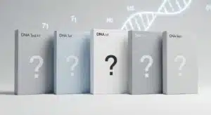
Genetics on Actinic Keratosis
Research on Actinic Keratosis where scientists have studied the genes involved in actinic keratosis (AK) and a type of skin cancer called cutaneous squamous cell carcinoma (cSCC) suggests that both conditions share many of the same genetic changes. However, the past studies often have looked at only specific candidate genes and had limitations
A few studies have looked at all the coding genes in AK using a method called whole-exome sequencing (WES), but those studies so far have had very few samples. Based on these studies it is now known that both AK and cSCC often have mutations in important genes like TP53, NOTCH1, NOTCH2, and FAT1. These genes help control how skin cells grow and stay healthy.
Overall, the study shows that AK and cSCC are very similar genetically. One important difference is with a cell communication pathway gene called TGFβ, which may play a key role in turning AK into skin cancer.
Can Actinic Keratosis Turn into Skin Cancer?
Most actinic keratoses (AKs) go away with treatment, and about 90% of people with AKs do not develop skin cancer. However, many cases of squamous cell carcinoma (SCC), a common type of skin cancer, start as AKs.
The risk of one AK turning into cancer is low, but if you have many AKs or ongoing sun damage, your chances of developing skin cancer are higher. The most serious risk associated with actinic keratosis is the potential development of cutaneous squamous cell carcinoma (cSCC). While the chance of a single AK turning into cSCC is low, people with multiple AK lesions (more than 10) have a 10–15% chance of developing SCC over time. Warning signs of possible cancerous changes include:
- Thickening or enlarging of the lesion
- Ulceration or bleeding
- Persistent tenderness or pain
- Rapid growth
Because AK indicates extensive sun damage, affected individuals are also at higher risk for other types of skin cancer, including basal cell carcinoma (BCC), melanoma, and rare forms like Merkel cell carcinoma.
How Is Actinic Keratosis Diagnosed?
Most actinic keratoses may be diagnosed by a dermatologist during a visual skin exam. If the spot looks unusual or they’re not sure, they may do a skin biopsy. This means they gently remove a small piece of the skin so it can be checked under a microscope.
An examination by a dermatologist helps confirm if a suspicious lesion is actinic keratosis or something more serious, like skin cancer. If there’s any uncertainty or suspicion of skin cancer, a dermoscopy or skin biopsy may be performed to examine the lesion more closely.
How Is Actinic Keratosis Treated?
There are several effective treatments available for actinic keratosis (AK). The choice of treatment depends on how many lesions you have and their appearance. Here are some common treatments:
- Cryotherapy: This is the most commonly used treatment. Liquid nitrogen is applied to freeze the AK, causing the damaged skin to blister and peel off. New, healthy skin then grows in its place. The procedure is quick and usually done in your provider’s office.
- Medicated Creams or Gels: For multiple or hard-to-see AKs, your healthcare provider may prescribe medicated creams such as those with fluorouracil, imiquimod, or diclofenac. These medicines work by removing damaged cells but can cause redness, peeling, and burning sensation during treatment.
- Excision: This involves numbing the skin, surgically cutting out the AK, and stitching the area closed. It is typically used for thicker or suspicious lesions and usually heals within two to three weeks.
- Chemical Peels: During a chemical peel, a medical-grade chemical solution is applied to the skin to remove the top damaged layer. This allows new, healthy skin to grow. The treated area may be sore or red for a few days after the procedure.
How Can You Protect Your Skin?
Preventing further sun damage is the key to avoiding AKs and reducing recurrence. Effective prevention includes:
- Using broad-spectrum sunscreen (SPF 30 or higher) daily, even in winter or cloudy weather; reapplied every 2 hours when outdoors.
- Avoiding sun exposure during peak UV hours (10 a.m. to 4 p.m.).
- Wearing sun-protective clothing, wide-brimmed hats, and UV-blocking sunglasses.
- Seeking shade when outside.
- Avoiding tanning beds and artificial UV light sources.
References: